A recent study from the lead scientific advisor on Coronavirus, has found that 82% of those studied with the virus developed Lymphopenia (abnormally low lymphocytes in the blood)
Dr Zhong Nanshan, who discovered the SARS coronavirus in 2003 is the leading advisor in investigating and managing the current coronavirus crisis.
Lymphopenia, which is a condition where a specific white blood cell that is part of the body’s first-line defence against diseases is dramatically reduced.
So, it may be very pertinent to look at the known nutritional elements which nourish the bodies own Lymphocyte making capability, as a means of focus should you feel the need to prepare, eat (or supplement) whilst your body is fighting this Virus.
Here is a strong, concise overview of such dietary elements:
Lymphocyte Nutritional Support:
Dietary Guidelines for a Better Lymphocyte Count You may want to know how to increase lymphocytes naturally. A healthy, nutrient-rich diet can go a long way toward boosting lymphocyte levels. This will provide your immune system with everything it needs to fight off viruses and bacteria that can potentially lead to low lymphocyte levels.
The following is a dietary guideline to follow to help your body improve its lymphocyte count.
Eat lots of lean protein: When the body doesn’t get enough protein, this leads to fewer white blood cells. As a result, you can increase lymphocyte production when you eat protein-rich foods such as grass-fed meats like poultry and beef, organic eggs, wild-caught fish and seafood, and legumes.
Avoid foods high in trans and saturated fats: These fats thicken lymphocytes; as such, reducing trans and saturated fat consumption can help improve immune system health. Avoid unhealthy fats such as margarine, fried foods, hydrogenated oils, and processed baked goods.
Consume healthy fats: Omega-3 fatty acids, on the other hand, will increase your lymphocyte count. Include omega-3 fatty acid foods such as avocado, ground flaxseed, hemp seeds, chia seeds, walnuts, sardines, albacore tuna, white fish, Alaskan salmon, herring, and Atlantic mackerel in your diet.
Eat foods high in beta-carotene: Beta-carotene helps boost lymphocyte production. Foods rich in beta-carotene include carrots, sweet potatoes, butternut squash, romaine lettuce, and spinach.
Eat zinc-rich foods: Zinc is needed to make lymphocytes. It also increases levels of NK cells and T cells, which strengthens your immune system. Foods high in zinc include oysters, asparagus, collard greens, spinach, broccoli, sesame seeds, and pumpkin seeds.
Consume foods high in vitamin C: Vitamin C is known to increase the production of white blood cells such as lymphocytes. Foods high in vitamin C include bell peppers, parsley, kale, oranges, raspberries, tomatoes, and celery.
Eat foods loaded with vitamin D: Not getting enough vitamin D can lower lymphocyte levels and weaken your immune system. Foods rich in vitamin D include organic eggs, raw milk, wild-caught salmon, sardines, mackerel, and tuna.
Eat foods high in vitamin E: Vitamin E supports production of NK cells and B cells. Foods rich in vitamin E include sunflower seeds, almonds, kale, spinach, olives, asparagus, and collard greens.
Eat selenium-rich foods: Selenium helps the body produce more white blood cells. Foods high in selenium include cod, shiitake mushrooms, salmon, tuna, eggs, oats, and broccoli.
Eat more garlic: Garlic is known to boost white blood cell production, which increases the number of NK cells. Purchase fresh, powdered, or dried garlic, and use it in your cooking daily.
Drink more green tea: Green tea compounds can boost immunity by fighting viruses that deplete white blood cells.
Low lymphocyte count, also known as lymphocytopenia, is a cause for concern because when lymphocytes (a type of white blood cell) are low, the body’s ability to repel infections is weakened.
ChooseLife : Of Course, I am always focused on the Acid/Alkaline balancing aspects in life, so this is not to discount the strong evidence that initial infectivity and severity it almost certain to be controlled by the hosts pH, if your tissue at the site of infection is below 7 you are much more likely to become infected, if 6 or lower it has been demonstrated in previous strains that the infectivity is 10x higher, Alkaline eating and supplementation is something I am very focused on for me and my family.
pH-Dependent Entry of Severe Acute Respiratory Syndrome Coronavirus Is Mediated by the Spike Glycoprotein and Enhanced by Dendritic Cell Transfer through DC-SIGN
June 2004
ABSTRACT
The severe acute respiratory syndrome coronavirus (SARS-CoV) synthesizes several putative viral envelope proteins, including the spike (S), membrane (M), and small envelope (E) glycoproteins. Although these proteins likely are essential for viral replication, their specific roles in SARS-CoV entry have not been defined. In this report, we show that the SARS-CoV S glycoprotein mediates viral entry through pH-dependent endocytosis. Further, we define its cellular tropism and demonstrate that virus transmission occurs through cell-mediated transfer by dendritic cells. The S glycoprotein was used successfully to pseudotype replication-defective retroviral and lentiviral vectors that readily infected Vero cells as well as primary pulmonary and renal epithelial cells from human, nonhuman primate, and, to a lesser extent, feline species. The tropism of this reporter virus was similar to that of wild-type, replication-competent SARS-CoV, and binding of purified S to susceptible target cells was demonstrated by flow cytometry. Although myeloid dendritic cells were able to interact with S and to bind virus, these cells could not be infected by SARS-CoV. However, these cells were able to transfer the virus to susceptible target cells through a synapse-like structure. Both cell-mediated infection and direct infection were inhibited by anti-S antisera, indicating that strategies directed toward this gene product are likely to confer a therapeutic benefit for antiviral drugs or the development of a SARS vaccine.
The severe acute respiratory syndrome coronavirus (SARS-CoV) is the likely cause of an acute infectious respiratory disorder identified in highly lethal outbreaks during the past year (10, 18, 21, 32, 40). Infection is characterized by acute flu-like symptoms that progress to a severe febrile respiratory illness with significant mortality. Coronaviruses, comprising a genus of the Coronaviridae family, are enveloped positive-strand RNA viruses. In general, coronaviruses cause respiratory and enteric diseases in humans and domestic animals (15, 20). Two previously known human coronaviruses caused only mild upper respiratory infections (15, 20). In contrast, a highly pathogenic, severe respiratory disease is caused by the SARS-CoV, especially in the elderly (44). Coronaviruses can be divided into three serologically distinct groups (15). Phylogenetically, SARS-CoV is not closely related to any of the three groups (26), though it is most similar to the group II coronaviruses (33, 36).
Although the organization of the SARS-CoV genome is related to that of animal coronaviruses, its genetic sequence is unique, and the structure and function of its gene products are not known. At least 14 open reading frames (ORFs) can be identified in its genome (26, 34, 36). Among these, the replicase/transcriptase genes are located in the 5′ portion of the genome. At its 3′ end, the four major structural proteins (S, M, N, and E) are made through different subgenomic RNAs. Based on comparison to animal coronaviruses, three structural gene products are predicted to be present on the viral envelope: the spike (S), membrane (M), and small envelope (E) proteins (20, 26, 34). The structure of the SARS-CoV envelope differs in some respects from that of other enveloped viruses, such as retroviruses and lentiviruses, many of which contain one viral envelope protein.
Envelope or spike proteins from enveloped viruses have been used to pseudotype retroviral and lentiviral vectors for functional and gene transfer studies (29, 35, 43, 45); however, whether coronavirus glycoproteins could pseudotype these viruses was unknown. Here we report that replication-defective retroviral (Moloney murine leukemia virus) and lentiviral (human immunodeficiency virus type 1 [HIV-1]) vectors can be pseudotyped with the SARS-CoV S protein, and the properties of S related to entry have been defined. Using these pseudoviruses, we were able to determine the relative contributions of SARS-CoV envelope proteins to viral entry and fusion and to examine the roles of these different viral envelope gene products with respect to entry, cell specificity, and potential inhibition of viral replication.
Pertinent Extract:
In contrast, influenza and Ebola viruses are prototypes for viruses that utilize a pH-dependent endocytotic pathway (43). To determine the pathway utilized by the SARS-CoV, the pH dependence of the SARS-CoV S-pseudotyped lentiviral vector was analyzed. Addition of ammonium chloride, which prevents acidification of the endosome, caused a dose-dependent reduction in viral entry (Fig. (Fig.1B,1B, left) at concentrations similar to those described for other pH-dependent viral glycoproteins (3, 11, 43). This effect was also observed with another inhibitor of endosomal acidification, bafilomycin, also in a dose-dependent fashion (Fig. (Fig.1B,1B, right).
Previous research from Meridian Institute Article :
Possible Relevance to SARS
The World Health Organization has concluded that SARS is produced by a new virulent strain of coronavirus. Specific research on the possible pH dependency of the SARS virus has not yet been done. It is well known that coronavirus infectivity is exquisitely sensitive to pH. For example, the MHV-A59 strain of coronavirus is quite stable at pH 6.0 (acidic) but becomes rapidly and irreversibly inactivated by brief treatment at pH 8.0 (alkaline). Human coronavirus strain 229E is maximally infective at pH 6.0. Infection of cells by murine coronavirus A59 at pH 6.0 (acidic) rather than pH 7.0 (neutral) yields a tenfold increase in the infectivity of the virus.
ChooseLife : If the strain of coronavirus responsible for SARS shares the pH characteristics of these other coronaviruses that are pH-dependent, this could be a valuable clue to effective prevention and treatment strategies for this potential epidemic. Perhaps keeping a balanced or slightly alkaline pH environment for the body’s tissues can provide viral protection or enhanced healing for SARS and common viral agents that cause respiratory infections.
Inter-related to this, is research on MUC5B, which has shown that those of lower pH, are much more prone to having inhibited mucous membrane formation:
“Moreover, we demonstrate that the conformation of these highly entangled linear polymers is sensitive to calcium concentration and changes in pH. In the presence of calcium (Ca2+, 10 mM) at pH 5.0, MUC5B adopted a compact conformation which was lost either upon removal of calcium with EGTA, or by increasing the pH to 7.4. These results suggest a pathway of mucin collapse to enable intracellular packaging and mechanisms driving mucin expansion following secretion. They also point to the importance of the tight control of calcium and pHduring different stages of mucin biosynthesis and secretion, and in the generation of correct mucus barrier properties. “
ChooseLife Related Thoughts :
The above shows that there are multiple potential protective methodologies in play, some people may feel a glass of cold water with 1/2 teaspoon of Sodium Bicarbonate every two hours on the first day may be effective (outlined at the bottom of this page), this is one method I would consider myself (Arm and Hammer or Bobs Mill being Aluminium free). Also small Sips of highly Alkaline Milk of Magnesia, every hour, may coat the upper respiratory regions fairly well and rapidly bring up the pH, out of the greater danger zones of lower pH < 6.5 (this is my go to for my kids with sniffles or worse), I would likely do this myself for this situation.
Personally I am going to use this outbreak as a good time to bring my own (and childrens) pH up, using methods as above, plus make some Moreless Alkalising Mineral Mixture, which both Alkalises and significantly raises the Calcium levels in the body but in a complexed form (pre-bonded to Molasses or Honey) which does not hamper the Mucous membrane process outlined above, which as shown above in the scientfic literature is exactly what our bodies need to be ready to either repel, or minimise the effects of such threats.
“The proven value of Bicarbonate of Soda as a therapeutic agent (from a letter to the Church and Dwight Company):
In 1918 and 1919 while fighting the Flu with the U.S. Public Health Service it was brought to my attention that rarely any one who had been thoroughly alkalinized with bicarbonate of soda contracted the disease, and those who did contract it, if alkalinized early, would invariably have mild attacks. I have since that time treated all cases of Cold, Influenza and LaGripe by first giving generous doses of Bicarbonate of Soda, and in many, many instances within 36 hours the symptoms would have entirely abated.
Further, within my own household, before Women’s Clubs and Parent-Teachers’ Association, I have advocated the use of soda as a preventative for ‘Colds’, with the result that now many reports are coming in stating that those who took ‘Soda’ were not affected, while nearly everyone around them had the ‘Flu’.
…An occasional three-day course of the Bicarbonate of Soda increases the alkalinity of the blood, assists elimination and increases the resisting power of the body to all Infectious Diseases…
Whenever taking a bicarbonate solution internally, the soda should be dissolved in cold water. In the event of a threatened attack we recommend the following treatment: During the first day take six doses of half a teaspoon of Bicarbonate of Soda in a glass of cool water, at about two hour intervals.”
New research finds close association between high aluminum content and amyloid-beta
Amsterdam, NL – A new study published in the Journal of Alzheimer’s Disease (JAD) supports a growing body of research that links human exposure to aluminum with Alzheimer’s disease (AD). Researchers found significant amounts of aluminum content in brain tissue from donors with familial AD. The study also found a high degree of co-location with the amyloid-beta protein, which leads to early onset of the disease.
“This is the second study confirming significantly high brain accumulation in familial Alzheimer’s disease, but it is the first to demonstrate an unequivocal association between the location of aluminum and amyloid-beta in the disease. It shows that aluminum and amyloid-beta are intimately woven in the neuropathology,” explained lead investigator Christopher Exley, PhD, Birchall Centre, Lennard-Jones Laboratories, Keele University, Staffordshire, UK.
An association between aluminum and amyloid-beta has been suggested for over 40 years. In an earlier study, brain tissue from donors in the United Kingdom diagnosed with familial AD showed significant accumulations of aluminum. To further understand this relationship, in the current study the researchers measured aluminum in the brain tissue of a cohort of Colombian donors with familial AD who shared a specific mutation. The mutation leads to elevated levels of amyloid-beta, early disease onset, and an aggressive disease etiology. The levels were compared with a control set of brain tissues from donors with no diagnosis of neuropathological disease. They also used aluminum-specific fluorescence microscopy imaging to investigate the relationship between aluminum and amyloid-beta in familial AD.
The results were striking. The aluminum content of the brain tissue from donors with the genetic mutation was universally high, with 42% of tissues having a level considered pathologically significant, and the levels were significantly higher than those in the control set. The imaging studies identified aluminum deposits in all brain tissues studied. They were predominantly co-located with amyloid-beta in senile plaques and occasionally in the brain vasculature. Aluminum was also found separately from amyloid-beta in intracellular compartments including glia and neuronal axons. The results strongly suggest that genetic predispositions known to increase amyloid-beta in brain tissue also predispose individuals to accumulate and retain aluminum in brain tissue.
“Compelling localization of aluminum with a central player in AD, amyloid-beta, strengthens the link of aluminum to the pathogenesis of AD,” commented George Perry, PhD, Professor of Biology, Semmes Distinguished University Chair in Neurobiology, University of Texas at San Antonio, and Editor-in-Chief of JAD.
“One could envisage increased amyloid-beta in brain tissue as a response to high levels of aluminum content, or that aluminum fosters the accumulation of amyloid-beta,” said Dr. Exley. “Either way, the new research confirms my resolve that within the normal lifespan of humans, there would not be any AD if there were no aluminum in the brain tissue. No aluminum, no AD.”
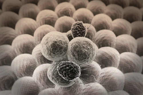
Amyloid-beta (green fluorescence) and aluminum (orange fluorescence) in senile plaque from brain tissue of a familial Alzheimer’s disease donor
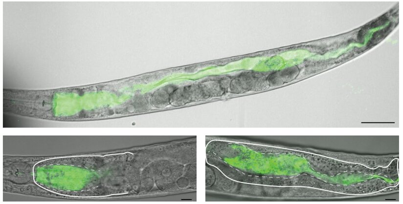
The investigation was performed using small, bacteria-eating organisms called Caenorhabditis elegans, here imaged using Oregon Green-labeled dextran and 748 laser-scanning confocal microscopy.
When food we’ve swallowed reaches our stomachs, it finds an acidic environment. The low pH in the stomach helps to begin digestion—and has been thought to kill the bacteria that hides in food that otherwise could harm our bodies.
However, recent work from the Ackley and Chandler labs in the Department of Molecular Biosciences at the University of Kansas runs counter to this idea, instead suggesting lower pH in the digestive tract may make some bacterial pathogens even more harmful.
Their findings, published in the peer-reviewed journal PLOS Pathogens, could have implications for addressing the crisis of antibiotic resistance in bacterial infections around the world.
The investigation was performed using small, bacteria-eating organisms called Caenorhabditis elegans.
“These wormlike animals are transparent, so we can watch things that happen inside them quite easily,” said co-author Brian Ackley, associate professor of molecular biosciences at KU. “Using pH-sensitive chemicals developed at KU, called Kansas Reds, we were able to monitor the pH inside the digestive system and watch what happens when they eat harmful bacteria, compared to nonharmful bacteria.”
According to the KU researchers, under normal conditions while feeding on healthy bacteria, C. elegans digestive tracts are moderately acidic compared to human stomachs. But these model species’ stomachs also show regional differences within the digestive tract. When they ingest pathogens, they neutralize the acidic environment.
This observation suggested the animals could discriminate between good and bad bacteria, and harmful bacteria prompted a less acidic digestive tract in C. elegans—a result that runs counter to what one might expect if the acidic environment was generated to kill bacteria.
To test this, the researchers used animals with mutations in genes that helped regulate the pH in their digestive tracts.
“When animals had a more acidic digestive system, they were more likely to be affected by pathogenic bacteria—again counter to what one might guess if acidity was useful in killing harmful bugs that might sneak into the body with food,” Ackley said. “Our lab teams were able to show the effect on the animals was specifically due to the pH by adding a base to buffer the digestive tract. We used bicarbonate, the same agent our bodies use to neutralize stomach contents when they pass into our intestines. Neutralizing the pH in the mutant animals reverted the accelerated infection by the pathogenic bacteria.”
The KU researcher said different species react differently when their bodies sense pathogenic bacteria—but some biological reactions are common to many animals.
“A general response involves the creation of chemicals, like hydrogen peroxide or hypochlorous acid—aka bleach—near the bacteria, and then having specialized immune cells eat the dying bacteria,” Ackley said. “To keep our bodies safe, the immune system only deploys these defenses when it’s sure it is being invaded. The work in C. elegans may suggest a way the body can have these defenses ready to go at a moment’s notice—that is, keep the chemical environment in a moderately acidic state where making those chemicals is difficult, then, upon infection, simply neutralize the environment to deploy the defenses.”
Ackley’s KU colleagues on the work were lead author Saida Benomar, Patrick Lansdon and Josephine R. Chandler of the Department of Molecular Biosciences, along with Aaron Bender of the Department of Medicinal Chemistry, and Blake R. Peterson of The Ohio State University.
The researchers believe there may be reasons to believe these systems could work similarly in people.
The genes they studied in C. elegans also exist in humans and control parts of the immune system. Further, research in other labs has shown occasions in humans where problems with regulating pH are associated with increased risk of infection. Moving forward, the researchers want to understand the mechanism at a deeper level.
“Our goal is to boost this natural defense system in people as a way to either avoid or reduce the use of antibiotics,” Ackley said. “Right now, our antibiotic use is unsustainable, and bacteria are evolving resistance at an alarming rate. If the system discovered in C. elegans is in fact still present in humans, it would suggest bacteria are much slower to adapt to this defensive strategy than they are to antibiotics.”
ChooseLife : Carey Reams stated that the production and regulation of stomach acid, is firstly regulated by the Liver, and, that primarily it’s production requires Calcium Salt in the form of Calcium Gluconate. Reams often proposed that a diet lower in Protein was better for health, so this may align to this concept, that those eating more meat or plant proteins may have higher stomach acid production, and, hence a generally lower pH. Then becoming more susceptible to these type of infections?
In 1984, as an undergraduate at the University of Stirling, Scotland and while carrying out my first piece of independent research, I watched for the first time a fish, a salmon parr, die from acute aluminium toxicity. The whole process took less than forty-eight hours. Within six hours, the fish showed signs of distress and its behaviour changed markedly. It proceeded to seek out the corners of the tank, pushing its head and body against the side of the tank. After twenty-four hours, it began to move randomly and chaotically around the tank before losing its orientation, slipping onto its back, taking a last gasp, before dying. I was left in no doubt about the toxicity of aluminium. I am recalling this event herein because there does seem to be significant complacency concerning the toxicity of aluminium.
An Aluminium Adjuvant is Acutely Toxic Too
In a recent post (https://www.hippocraticpost.com/pharmacy-drugs/the-toxicity-of-aluminium-adjuvants/) I explained why a single injection of a vaccine that includes an aluminium adjuvant is, akin to the salmon above, also an acute exposure to aluminium. It is acute because the total concentration of aluminium in the immediate vicinity of the injection site is extremely high, in the case of a single dose of Infanrix Hexa vaccine, approximately 8000 times higher than is required to kill a salmon parr within forty-eight hours. Even allowing for some dilution of the injected aluminium adjuvant into body fluids bathing and innervating the tissues surrounding the injection site the total concentration of aluminium in a vaccine is sufficient to cause cell death within hours and perhaps minutes of receiving the injection. This is the definition of an acute response, death (cells or whole organism) within a short period of exposure to a toxin. It is a necrotic form of cell death. It initiates an inflammatory response (redness at the injection site). This inflammation drives and perhaps accelerates the subsequent immune response (https://www.hippocraticpost.com/infection-disease/safety-concerns-aluminium-adjuvants/).
A number of mechanisms bring about remediation of acute aluminium toxicity at a vaccine injection site. These are chemical, physical and biological. The toxic free metal ion, Al3+, forms soluble and insoluble complexes with myriad biological molecules while particles of aluminium adjuvant and other insoluble aluminium compounds are taken up by cells infiltrating the vaccine injection site. All of these processes act to reduce the acute toxicity of aluminium at the injection site by lowering the immediate concentration of toxic Al3+. These remedial processes act to secure aluminium in a number of different compartments. All are systemic and all are potential sources of biologically reactive aluminium to the rest of the body. Many chemical compartments where aluminium is bound in myriad different complexes including simple organic moieties like citrate or more complex proteins like the iron transport protein transferrin promote the transport of aluminium away from the injection site.
These processes can be envisaged as continuous passive diffusion of soluble aluminium away from the injection site. The majority of injected aluminium adjuvant is particulate in the first instance and actively taken up, literally eaten, by a number of different cells infiltrating the injection site. Some particles of aluminium adjuvant are taken up by macrophages and thereafter they are retained at or close to the injection site as a granuloma. Generally, these collections of macrophages are considered as benign ‘cancers’ though such descriptions have been coined for situations where the cellular cargo is not aluminium. For example, macrophagic myofasciitis or MMF is a disease, first described by Romain Gherardi in Paris, in which aluminium-rich granulomas at vaccine injection sites are implicated in disease aetiology. Other cells heavily laden with aluminium do not remain close to the injection site and carry their cargo well beyond where the vaccine is administered, for example visiting local lymph nodes as early stops on their travels. Evidence is mounting that these cells may transport aluminium into brain tissue using both lymph and blood as access routes. Perhaps most worrying, evidence of transport of aluminium into brain tissue across the blood-brain barrier and meninges has been shown in autism (https://www.hippocraticpost.com/infection-disease/aluminium-and-autism/).
An Aluminium Adjuvant is a Significant Exposure to Aluminium
With reference to my recent post, an aluminium adjuvant in a vaccine is an acute exposure to aluminium at the vaccine injection site. However, the aluminium content of a single vaccine also represents a significant exposure to aluminium in an infant. For example, the injection of a single dose of Infanrix Hexa into an infant is equivalent to 164 times the daily dose of aluminium in breast milk feeding. Even allowing for an unrealistically high proportion of aluminium being retained in a granuloma at the vaccine injection site (say, for example 40% of the injected aluminium) the daily dose of aluminium in Infanrix Hexa is 100 times higher than an infant receives in breast-feeding. This is a high exposure to aluminium and inevitably results in aluminium being retained in an infant’s tissues, including the infant brain. This is why we must not be complacent about the use of aluminium adjuvants in vaccines. It is why there should be regulations based upon the toxicity of aluminium that govern how much aluminium is allowed in a single vaccine. This limit should be used to give clear unequivocal advice on the number of vaccines that include an aluminium adjuvant that can be given within a specified period. Aluminium is only toxic (as opposed to essential) in the human body and so we should always take every possible step to reduce exposure and ultimately the body burden of aluminium. Infants, due to increased gastrointestinal absorption, reduced urinary excretion and a developing blood-brain-barrier, are uniquely vulnerable to aluminium. We need to protect them from their future now.
Professor in Bioinorganic Chemistry Keele University Honorary Professor, UHI Millennium Institute Group Leader – Bioinorganic Chemistry Laboratory at Keele
Americans are undeniably great consumers of added sugar. The average person’s intake is over 126 grams of sugar daily which is way too much. Sugar per se is not harmful but too much of it is detrimental to health. High sugar consumption has long been linked to weight gain, and the development of obesity and lifestyle diseases such as heart disease and diabetes. More recent studies show that sugar can also induce memory problems and neuroinflammation.
A study conducted at the University of Southern California (USC) revealed that intakse of high fructose corn syrup (HFCS) at levels similar to those in commonly available sugary beverages can trigger memory problems and brain inflammation. In the study, adolescent rats consuming high quantities of HFCS were subsequently found to have impaired hippocampal-dependent spatial learning and memory when subjected to the Barne’s Maze test. Rats consuming a sucrose solution experienced moderate learning impairment. Adult rats given either HFCS or sucrose did not show any problems with spatial learning, glucose tolerance or neuroinflammatory markers. According to Scott Kanoski, one of the proponents of the research, the brain is vulnerable to dietary influences during critical periods of development, like adolescence. He further explained that a diet high in added sugar can not only cause weight gain and metabolic problems, but also impair brain function and cognitive ability.
This is supported by an earlier study at the University of California in Los Angeles (UCLA) that tested the effects of fructose and omega-3 fatty acid consumption among rats. Two groups of rats were initially trained to find their way out of a maze. The results showed that long-term consumption of a high fructose diet by the rats negatively affected their cognitive function and ability to recall while omega 3 fatty acids were able to counteract those effects. After six weeks, the rats that consumed fructose forgot the previously learned escape route and developed signs of insulin resistance. The impaired learning and memory problems exhibited by this group of rats was attributed to insulin’s inability to regulate how cells use and store sugar.
Sucrose and high fructose corn syrup are liquid sweeteners often added to processed foods and common beverages such as soft drinks and juices. These added sugars are quickly absorbed in the blood where it lowers the production of brain-derived neurotrophic factor (BDNF), a substance known to influence the formation of new memories and regulate learning. Individuals with impaired glucose tolerance were found to have low levels of this chemical. Low levels of BDNF have also been associated with depression and dementia.
So before you drink your favorite juice or munch on a delectable dessert, remember that added sugars from such beverage and food items are likely culprits for health complications including poor memory and cognitive damage. Avoiding foods with added sugars will help you stay fit and keep your mind healthy.
The polymeric mucin MUC5B provides the structural and functional framework of respiratory mucus, conferring both viscoelastic and antimicrobial properties onto this vital protective barrier. Whilst it is established that MUC5B forms disulfide-linked linear polymers, how this relates to their packaging in secretory granules, and their molecular form in mucus remain to be fully elucidated. Moreover, the role of the central heavily O-glycosylated mucin domains in MUC5B conformation is incompletely described. Here we have completed a detailed structural analysis on native MUC5B polymers purified from saliva and subsequently investigated how MUC5B conformation is affected by changes in calcium concentration and pH, factors important for mucin intragranular packaging and post-secretory expansion. The results identify that MUC5B has a beaded structure repeating along the polymer axis and suggest that these repeating motifs arise from distinct glycosylation patterns. Moreover, we demonstrate that the conformation of these highly entangled linear polymers is sensitive to calcium concentration and changes in pH. In the presence of calcium (Ca2+, 10 mM) at pH 5.0, MUC5B adopted a compact conformation which was lost either upon removal of calcium with EGTA, or by increasing the pH to 7.4. These results suggest a pathway of mucin collapse to enable intracellular packaging and mechanisms driving mucin expansion following secretion. They also point to the importance of the tight control of calcium and pH during different stages of mucin biosynthesis and secretion, and in the generation of correct mucus barrier properties.