Andrew Benner; Aakash K. Patel; Karampal Singh; Anterpreet Dua.
Introduction
Oxygen (O2) competitively and reversibly binds to hemoglobin, with certain changes within the environment altering the affinity in which this relationship occurs. The sigmoidal shape of the oxygen dissociation curve illustrates hemoglobin’s propensity for positive cooperativity, as hemoglobin undergoes conformational changes to increase its affinity for oxygen as molecules progressively bind to each of its four available binding sites. The Bohr effect describes hemoglobin’s lower affinity for oxygen secondary to increases in the partial pressure of carbon dioxide and/or decreased blood pH. This lower affinity, in turn, enhances the unloading of oxygen into tissues to meet the oxygen demand of the tissue.[1]
Issues of Concern
Increases in PCO2 and Decreases in pH
Through the biochemical reactions necessary for cellular respiration, increases in metabolic activity within tissues result in the production of carbon dioxide (CO2) as a metabolic waste product. This increase in tissue PCO2 leads to an increase in hydrogen ion (H+) concentration, represented as a decrease in pH as the environment undergoes the process of acidosis. These effects decrease hemoglobin’s affinity for oxygen, weakening its binding capacity and increasing the likelihood of dissociation; this is represented as a rightward shift of the hemoglobin dissociation curve, as hemoglobin unloads oxygen from its binding sites at higher partial pressures of oxygen. Specifically, it is the association of protons (H+ ions) with the amino acids in hemoglobin that cause a conformational change in protein folding, ultimately reducing the affinity of the binding sites for oxygen molecules. This configuration shift of hemoglobin under the influence of protons is classified as the taut (T) form.
Hemoglobin exists in 2 forms, the taut form (T) and the relaxed form (R). This structural change to the taut form leads to low-affinity hemoglobin, whereas the relaxed form leads to a high-affinity form of hemoglobin with respect to oxygen binding. In the lungs, the highly saturated oxygen environment can overcome the lower affinity T-form of hemoglobin, effectively binding despite disadvantageous binding capacity. During this process, initial O2 binding induces an alteration in hemoglobin from the taut to relaxed form, dissociating H+ protons and progressively increasing hemoglobin’s affinity for oxygen at each of the remaining binding sites through positive cooperativity. Under the influence of acidic environments, hemoglobin has a propensity for undergoing the reverse of this conformational change, releasing oxygen in favor of the attachment of H+ protons as hemoglobin shifts from the higher oxygen affinity relaxed form to the lower oxygen affinity taut form.
Overall, this relationship can be quantified by an increase in the P50,as 50% hemoglobin oxygen saturation is achieved at higher-than-normal values of pO2 compared to the accepted normal P50 of 27 mmHg. This results in greater unloading of oxygen in the presence of the acidic environments surrounding body tissues as a result of cellular respiration.[2]
Cellular Level
Through the enzyme carbonic anhydrase, the carbon dioxide and water released as byproducts from cellular respiration are converted to carbonic acid (H2CO3). In pursuing biochemical equilibrium, carbonic acid partially and reversibly dissociates into hydrogen ions and its conjugate base, bicarbonate (HCO3). This release of hydrogen ions increases the available concentration of H+ ions within the blood, effectively decreasing the pH of the environment. Due to the reversibility of this reaction, the resulting bicarbonate conjugate base form of carbonic acid indirectly represents the majority of the blood carbon dioxide (CO2) content (70%).[3]
CO2 + H2O <-> H2CO3 <-> H+ + HCO3-
This process usually takes place in peripheral tissues, as the desired effect is to unload oxygen into these tissues and load oxygen in the lungs. To limit the decrease in pH of the environment surrounding peripheral tissues, hemoglobin serves as a buffering agent by releasing its oxygen molecules in favor of binding H+ ions. Additionally, the increased bicarbonate molecules move down their concentration gradient, diffusing out of the red blood cell, exchanging chlorine ions into the red blood cell to maintain electrical neutrality. This buffering process is known as the Haldane effect. In the setting of lung alveoli, the less acidic and highly oxygenated environment favors the dissociation of the scavenged H+ protons from hemoglobin in exchange for oxygen binding. The effect of this relatively increased pH environment and its effect on hemoglobin oxygen affinity is graphically represented as a left shift in the oxy-hemoglobin dissociation curve as the P50 effectively decreases, resulting in greater attachment of oxygen to hemoglobin.[4]
Related Testing
The measurement of the oxygen and carbon dioxide content in the blood, in addition to acid-base status in the form of pH level, is quantifiable through an arterial blood gas (ABG) analysis. As a result, the global interpretation of all available data within an ABG provides an approximation of the body’s ventilation and metabolism efforts. Based on the PaCO2 on the blood gas, clinicians can get a sense of the amount of CO2 retention and the effect it may have on the Bohr effect, and ultimately oxygen delivery to body tissues. The accepted “normal” PaCO2 concentration is commonly described as 40 mm Hg, with hypercapnia and hypocapnia defined as a PaCO2 greater than 45 and less than 35 mmHg, respectively. In the clinical circumstance of PaCO2 greater than 45 mmHg combined with a PaO2 less than 60 mm Hg, the patient may be experiencing hypercapnic respiratory failure with an ensuing right shift in the oxygen dissociation curve to increase oxygen delivery.
Pathophysiology
Through the Bohr effect, more oxygen is released to those tissues with higher carbon dioxide concentrations. The sensitivity to these effects can be suppressed in chronic diseases, leading to decreased oxygenation of peripheral tissues. Chronic conditions such as asthma, cystic fibrosis, or even diabetes mellitus can lead to a chronic state of hyperventilation to maintain adequate tissue oxygenation. These states can have ventilation of up to 15 L per minute compared to the average normal minute ventilation of 6 L per minute. This hyperventilation minimizes the potential of the Bohr effect through excess exhalation of carbon dioxide resulting in hypocapnia, causing a left shift in the oxygen dissociation and unnecessarily increased oxygen-hemoglobin binding affinity with impaired oxygen release to peripheral tissues, including our most vital organs (brain, heart, liver, kidney). Thus, the Bohr effect is essential in maximizing oxygen transport capabilities of hemoglobin and functionally dynamic oxygen-binding/release secondary to carbon dioxide equilibrium. [5]
Carbon Monoxide Effect
While the presence of carbon dioxide leads to the greater unloading of oxygen, carbon monoxide has the opposite effect. Carbon monoxide (CO) has a 200-times greater affinity for hemoglobin than oxygen, out-competing oxygen for available binding sites in a nearly irreversible fashion (reversible, but very minimally). Carbon monoxide further decreases oxygen delivery through the stabilization of hemoglobin in the R-form. Counter-intuitively, although this facilitates oxygen loading to the remaining binding sites, hemoglobin becomes resistant to environmental influences that would normally encourage conformational changes into taut-form, limiting the potential for unloading of oxygen. Under the influence of carbon monoxide, the oxy-hemoglobin dissociation curve significantly shifts left in addition to the reduction of the sigmoidal curve shape as a result of blunted positive cooperativity response of hemoglobin. In the presence of significant carbon monoxide inhalation, tissue hypoxia occurs despite normal pO2 levels, as carbon monoxide competitively binds hemoglobin while inhibiting the release of oxygen from the remaining binding sites. Carbon monoxide poisoning is treated with hyperbaric oxygen therapy, delivering 100% O2 at increased atmospheric pressures to facilitate hemoglobin oxygen binding in the presence of highly competitive carbon monoxide.[6]
Double Bohr Effect
The Double Bohr effect is seen in the fetus. In the placenta, maternal and fetal circulation meets. The umbilical arteries carry de-oxygenated blood with high CO2 content from the fetus to the placenta. In the placenta, CO2 from fetal blood diffuses into maternal blood down its concentration gradient. As CO2 content of fetal blood decrease, this makes fetal blood relatively alkaline and shift the oxygen dissociation curve toward left, facilitating more oxygen uptake by fetal Hb.On the maternal side, this CO2 diffusion from the fetal side makes maternal blood in the placenta more acidic. This shifts ODC towards the right and more oxygen is released from maternal Hb. Thus in the placenta, the Bohr effect occurs twice, one on the fetal side and another on the maternal side. This is known as the double Bohr effect. The clinical significance of the double Bohr effect is that it facilitating oxygen transfer across the placenta from mother to fetus and thus increase fetal oxygenation. Fetal Hb also has more affinity for oxygen than adult Hb. P50 ( partial pressure at which the hemoglobin molecule is half saturated with O2) for fetal Hb is 19 whereas P50 of adult Hb is 27. This Low P50 of fetal Hb also favors more oxygen transfer to the fetus.
Clinical Significance
The Bohr effect describes red blood cells’ ability to adapt to changes in the biochemical environment, maximizing hemoglobin-oxygen binding capacity in the lungs while simultaneously optimizing oxygen delivery to tissues with the greatest demand. The Bohr effect maintains significant clinical relevance within the field of Anesthesiology, as it directly influences patient outcomes throughout the perioperative process. Whether through hypo or hyperventilation, the alterations in carbon dioxide content and acid-base status results in shifts in the oxy-hemoglobin dissociation curve, either amplifying or dampening the magnitude of the Bohr Effect regarding hemoglobin re-oxygenation at the alveoli and delivery/release at peripheral tissues.
So, the Russian study says as a headline, we’re all good, no significance found between Test and Control. However the details say, test subject Wistar Rats ate twice as much, gained no extra weight, then autopsy found intestinal inflammation.
Strange, to see this year, in myself, a young loved one and several others heightened intenstinal discomfort. The discomfort felt by myself felt exactly like that experienced in 2019 when 5G initially rolled out.
So what? After watching this video, check coverage maps and see that a new form of 5G is being rolled out “Supersonic 5G”, also marketed as “Ultra 5G”. A far more powerful 5G, what could go wrong? When checking, although we have no 5G in my house and live very remotely in Rural Dorset, there is still saturated coverage from EE, strong coverage from Vodafone, didn’t want to check any further, we are basically being bombarded why? So we can download a film in seconds rather than minutes with 4G?
Ultra bad for us, more like? Truly wish we could turn it all off. Personally we have lots of Orgonite around windows, noticed these past 5G years how Birds continually fly up to my windows, Robins have been in my house, Birds hit the Windows. The shed where I make Orgonite had a Bat nested in it, which entered as I had poured some Orgonite.
Just last night a Cat was at my lounge window, pawing at the Window to come in. Animals, they know?
September 2019 – Robin in my Bedroom, right at a time my insides were tightening right up, intuitively I had placed lots of pieces around my Bed/Bedroom as I suffered nightmares and digestive troubles.Something compelled me to make lots of Orgonite in the Summer of 2019. After years of inactivity. Offered this Ruby, Apatite, Tourmaline, 24ct Gold and 6ct White Gold piece to a Rinpoche who I had been enjoying learning from, but he didn’t accept the gift offer, so it sits under my bed.June 2022 – Unknown breed of Bird perching in my spare room/Orgonite Room Velux.June 2022 – Robin again in House, flying around lounge, then upstairs resting (or sending a message? Given my digestive challenges since 5G went live and todays post about science finding intestinal inflammation, a profound message?) on my Toilet seat.
These pictures, as I look back through them, show that the Birds were very active in periods a lot of Orgonite was being made and around, a refuge?
The same month (June 2022) as the Robin came in a concerted effort was made, to make lots of new Orgonite. Similarly in 2019 something inside compelled me to make lots for the first time in many years. This photo showed the swirl of Qi from a piece setting in its Martini Glass Mould, White Gold and Aquamarine core producing this visually perceptible picture.August 2022 lots of Gold and Heart stones (Rose Quartz and Morganite) were being made. Plus Throat (Blue – Aquamarine and Apatite). This piece was the nicest feeling, so it resides upon my Girls windowsill at their mums house.October 2022 a Bat takes refuge, they of course travel using the Earths Magnetic Field. The shed at that time was my Orgonite making area, after a Bees nest took over my spare room (previous Orgonite making room).The very same month I had been making that late summers Orgonite, the Bat flew in to take refuge?Many were made, many gifted to friends, people encountered, or buried in the Earth to gift our Earths beleaguered Spirits.
This year, multiple birds have hit closed windows, such that when I cut back my Hawthorne Bush between two lounge windows, two dead birds were found (who likely killed themselves hitting my Windows, trying to refuge from the Electromagnetic Storm mankind in inflicting upon itself and our creatures?).
Over ninety years ago, on November 8, 1845, Michael Faraday investigated the magnetic properties of dried blood and made a note “Must try recent fluid blood.” If he had determined the magnetic susceptibilities of arterial and venous blood, he would have found them to differ by a large amount (as much as twenty per cent for completely oxygenated and completely deoxygenated blood); this discovery without doubt would have excited much interest and would have influenced appreciably the course of research on blood and hemoglobin.1
Continuing our investigations of the magnetic properties and structure of hemoglobin and related substances,2 we have found oxyhemoglobin and carbonmonoxyhemoglobin to contain no unpaired electrons, and ferrohemoglobin (hemoglobin itself) to contain four unpaired electrons per heme. The description of our experiments and the interpretation and discussion of the results are given below.
Note on Nomenclature
The current nomenclature of hemoglobin and related substances was formulated at a time when precise information about the chemical composition and structure of the substances was not available. Now that some progress has been made in gathering this information, especially in regard to chemical composition, it is possible to revise the nomenclature in such a way as to make the names of substances more descriptive than the older names, without introducing any radical changes. In formulating the following set of names we have profited by the continued advice of Dr. Alfred E. Mirsky.
The names whose use we advocate are given below, followed in some cases by acceptable synonyms. The expressions in parentheses are those whose use we consider to be undesirable.
Heme: an iron-porphyrin complex (generic term, used for either ferroheme or ferriheme).
Ferroheme (reduced heme): a complex of ferrous iron and a porphyrin.
Ferriheme (oxidized heme): a complex of ferric iron and a porphyrin.
Ferriheme chloride, hemin: a compound of ferriheme and chloride ion.
Ferriheme hydroxide, hematin: a compound of ferriheme and hydroxyl ion.
Ferrohemochromogen, hemochromogen: a complex of ferroheme and another substance, or two other substances, having the characteristic hemochromogen spectrum and involving covalent bonds from the iron atom to the porphyrin nitrogen atoms and the attached groups.2 Individual hemochromogens may be designated by specifying the attached groups, as globin hemochromogen (ferroheme and denatured globin), dicyanide hemochromogen, dipyridine hemochromogen, carbonmonoxyhemochromogen, pyridine carbonmonoxyhemochromogen, etc.
Ferrihemochromogen (parahematin): a compound of ferriheme and another substance or two other substances, involving covalent bonds from the iron atom to the porphyrin nitrogen atoms and the attached groups.3
Hemoglobin: a conjugated protein containing heme and native globin (generic term, used for both ferrohemoglobin and ferrihemoglobin and also for closely related substances); specifically, ferrohemoglobin.
Ferrohemoglobin, hemoglobin (reduced hemoglobin): a conjugated protein formed by combination of ferroheme and native protein.
Oxyhemoglobin: a compound of ferrohemoglobin and oxygen.
Carbonmonoxyhemoglobin, carbon monoxide hemoglobin (carboxyhemoglobin): a compound of ferrohemoglobin and carbon monoxide.
Ferrihemoglobin (methemoglobin): a conjugated protein formed by combination of ferriheme and native globin.3
Carbonmonoxyhemoglobin
The magnetic measurements are described in the experimental part below. The carbonmonoxyhemoglobin molecule is found to have zero magnetic moment, and hence to contain no unpaired electrons. This is to be interpreted as showing that at least two 3d orbitals of each ferrous iron atom are involved in covalent bond formation, the atom presumably forming six octahedral d2sp3 bonds, four to the porphyrin nitrogen atoms, one to an atom (probably nitrogen) of the globin, and one to the carbon monoxide molecule:
In view of the discovery of Brockway and Cross4 that the nickel-carbon bond in nickel carbonyl has a large amount of double bond character, we may well expect this to be the case for the iron-carbon bond in carbonmonoxyhemoglobin also, the double bond being formed with the use of a pair of electrons conventionally assigned to the iron atom as 3d electrons. To carbonmonoxyhemoglobin there would then be ascribed the resonating structure:
in which the dashes represent shared electron pairs and the dots unshared electrons.
Oxyhemoglobin
The molecule of oxyhemoglobin, like that of carbonmonoxyhemoglobin, is found to have zero magnetic moment and to contain no unpaired electrons. Each iron atom is accordingly attached to the four porphyrin nitrogen atoms, the globin molecule, and the oxygen molecule by covalent bonds.
The free oxygen molecule in its normal state () contains two unpaired electrons. It might well have been expected, in view of the ease with which oxygen is attached to and detached from hemoglobin, that the oxygen molecule in oxyhemoglobin would retain these unpaired electrons, a pair of σ electrons of one oxygen atom, unshared in the free molecule, being used for the formation of the bond to hemoglobin:
However, this is shown not to be so by the magnetic data, there being no unpaired electrons in oxyhemoglobin. The oxygen molecule undergoes a profound change in electronic structure on combination with hemoglobin.
Of the structures of oxyhemoglobin compatible with the magnetic data, the most probable is the resonating structure analogous to that of carbonmonoxyhemoglobin:
The great similarity in properties of oxyhemoglobin and carbonmonoxyhemoglobin provides strong support for this structure. The structure in which each of the two oxygen atoms (connected with one another by a single bond) is attached to the iron atom by a single bond is rendered improbable by the strain involved in the three-membered ring.
Ferrohemoglobin
In contrast to oxyhemoglobin and carbonmonoxyhemoglobin, hemoglobin itself contains unpaired electrons, its magnetic susceptibility showing the presence of a pronounced paramagnetic contribution. The interpretation of the magnetic data can be made only in conjunction with a discussion of the nature and magnitude of the mutual interactions of the four hemes in the molecule.5 One possibility is that the heme-heme interaction is sufficiently strong to couple the moments of all electrons in the molecule into a resultant moment, with the same value for all molecules. The magnetic data interpreted in this way lead to the value μ= 10.92 Bohr magnetons for the moment of the molecule. We reject this possibility on the following grounds. (1) The heme-heme interaction energy, as evaluated from the oxygen equilibrium data,5 is hardly large enough to overcome the entropy advantage of independent heme moments. (2) The value 10.92 for the moment is not far from that (8.94) for eight unpaired electrons, two per heme, with parallel spins; however, it is about 22% larger, and this difference could be accounted for only as a surprisingly large contribution of orbital moment. (3) On this basis the magnetic susceptibility of partially oxygenated hemoglobin solutions would show large deviations from a linear dependence on the amount of uncombined heme; we have found large deviations not to occur. (These experiments will be described in a later paper.)
The other simple possibility, which we believe to be approximated in reality, is that the magnetic moments of the four hemes orient themselves in the applied magnetic field independently of one another. With the calculations made on this assumption, the experimental data lead to the value μ = 5.46 Bohr magnetons for the effective moment per heme. This shows that there are present in each heme four impaired electrons, and that consequently the iron atom is not attached to the four porphyrin nitrogen atoms and the globin molecule by covalent bonds, but is present as a ferrous ion, the bonds to the neighboring atoms being essentially ionic bonds.
The resultant spin moment for four unpaired electrons is 4.90 magnetons. In compounds containing ferrous ion values of 4.9 to 5.4 are observed, the increase over the spin moment arising from a small orbital contribution. Complexes of ferrous iron with substances containing nitrogen (hydrazine, etc.) give values in the lower part of this range, the quenching of orbital moment being nearly complete.6 It does not seem probable that the high value for ferrohemoglobin is to be accounted for as due to orbital moment, since the porphyrin nitrogen atoms should have a strong quenching effect on the orbital moment. We interpret this high value instead as due to a heme-heme interaction which tends to stabilize states with parallel heme moments relative to those with opposed heme moments, the oxygen-equilibrium value of the heme-heme interaction energy being of the order of magnitude required for this interpretation.
It is interesting and surprising that the hemoglobin molecule undergoes such an extreme structural change on the addition of oxygen or carbon monoxide; in the ferrohemoglobin molecule there are sixteen unpaired electrons and the bonds to iron are ionic, while in oxyhemoglobin and carbonmonoxyhemoglobin there are no unpaired electrons and the bonds are covalent. The change from ionic bonds to covalent bonds also occurs on formation of hemochromogen from ferroheme. Such a difference in bond type in very closely related substances has been observed so far only in hemoglobin derivatives.
It is not yet possible to discuss the significance of these structural differences in detail, but they are without doubt closely related to and in a sense responsible for the characteristic properties of hemoglobin. For example, the change in multiplicity of the system oxygen molecule–heme in hemoglobin on formation of oxyhemoglobin need be only as great as two (from the triplet corresponding to the opposed oxygen molecule triplet and ferroheme quintet to the singlet of oxyheme), whereas the change in multiplicity on formation of carbonmonoxyhemoglobin is four; in view of the infrequency of transitions involving a change in multiplicity, we might accordingly anticipate that the reactions of hemoglobin with carbon monoxide would be slower than those with oxygen, in agreement with observation. The change in multiplicity may be related also to the photochemical reactivity of carbonmonoxyhemoglobin. The difference in bond type in hemoglobin and its compounds is probably connected with the preferential affinity of hemoglobin for oxygen and carbon monoxide in contrast to other substances. Further experimental information is needed before these questions can be discussed in detail.
Experiments
Solutions: Defibrinated bovine blood (provided through the courteous coӧperation of Cornelius Bros., Ltd.) was used as the source of material. Preparations A and B consisted of whole blood, collected and separately oxygenated by rotating 20 minutes in air in a large open vessel, and then packed in ice and used as soon as possible. For preparations C and D oxygenated blood was centrifuged, and the corpuscles washed three times with equal volumes of physiological sodium chloride. Ether was used to hemolyze the collected corpuscles, the stromata-emulsions were separated by centrifuging, and the dissolved ether removed from the oxyhemoglobin solutions by a current of air. The solutions were kept on ice until used.
Analyses were made for oxygen content in a Van Slyke-Neill constant-volume blood gas apparatus. The transfer pipet was calibrated for content and retention on the walls of whole blood or concentrated oxyhemoglobin solution corresponding to conditions of use; the gas pipet was also calibrated for volume. Correction was made for dissolved oxygen on the assumption that the quantity dissolved is proportional to the water present in the solution.
Corrected results of analyses: Blood A: 100 ml. combine with 20.20 ml. O2 S.T.P.; formality of heme-iron, 0.00902. Blood B: 100 ml. combine with 20.59 ml. O2 S.T.P.; formality of heme-iron, 0.00919. Solution C: 100 ml. combine with 37.15 ml. O2 S.T.P.; formality of heme-iron, 0.01658. Solution D: 100 ml. combine with 41.26 ml. O2 S.T.P.; formality of heme-iron, 0.01841.
Apparatus: The apparatus for magnetic susceptibility determinations has already been described.2 All hemoglobin solutions were measured against water in a tube of about 18 mm. internal diameter. Fields of 7640 and 8830 gauss were used, the forces being reported as average Δw (in milligrams) for the former. A small correction to the observed Δw has been applied for blank on the tube, so that reported forces are for solution against pure water. Solutions were measured at approximately 20°C.
Calibration of field and tube with water against air: Δw = −49.59. (For hemochromogen and 6NNaOH the tube with Δw = −45.40 for water against air was used.)
Carbonmonoxyhemoglobin
Samples of blood A equilibrated with CO by rotation of 50 ml. in a liter tonometer filled with pure carbon monoxide: Δw = −0.56, −0.60, −0.76, −0.61, average −0.63. Samples of blood Bequilibrated with CO: Δw = −0.84, −1.03, −0.80, −0.86, average −0.88. Samples of solution Cequilibrated with CO: Δw = −0.28, −0.68, average −0.48. Samples of solution D equilibrated with CO: Δw = −0.36, −0.45, average, −0.41. (Completeness of saturation with carbon monoxide was generally tested by adding Na2S2O4.2H2O to the magnetic tube and measuring the increase in susceptibility due to formation of hemoglobin.)
We have established in the previous paper2 the presence of no unpaired electrons in globin hemochromogen and dicyanide hemochromogen. For globin hemochromogen made by denaturing 32 ml. of whole blood with 10 ml. of 6N NaOH after reduction of the heme: average Δw = −1.71; for dicyanide hemochromogen prepared in a similar manner: average Δw = −1.53; average for the two, Δw = −1.62. Measurement of the 6N NaOH against water in the same tube gives Δw = −4.88, −4.95. Assuming the additivity of atomic diamagnetism (Wiedemann’s rule), whole blood without paramagnetic constituent should give Δw = −0.58 in the tube used for the hemoglobin series. This value is in satisfactory agreement with the Δw values given above. The calculated value for blood with two unpaired electrons per heme, and independent hemes, is Δw = +1.52, for four, Δw = +5.69; the calculated values for the hemoglobin solutions are about twice as great.
Conclusion: carbonmonoxyhemoglobin contains no unpaired electrons.
Oxyhemoglobin
Samples of blood A: Δw = −0.65, −0.40, −0.44, average, −0.50. Blood B: Δw = −0.58, −0.62, −0.62, average, −0.61. Solution C: Δw = −0.44, −0.55, −0.50, −0.50, average, −0.50. Solution D: Δw = −0.38, −0.36, average, −0.37. Oxyhemoglobin relative to carbonmonoxyhemoglobin: A, +0.13; B, +0.27; C, +0.02; D, +0.04; calculated for two unpaired electrons on oxyhemoglobin: A, +2.03; B, +2.07; C, +3.74; D, +4.11.
Conclusion: oxyhemoglobin contains no unpaired electrons.
Hemoglobin
35 ml. of blood A reduced in differential tube by addition of from 0.4 to 1.0 g. Na2S204.2H20: Δw= +7.32, 6.85, 6.98, 6.97, average, +7.03. Taking the mean of the oxy- and carbonmonoxyhemoglobin values (−0.57) for Δw of diamagnetism of hemoglobin, the change on removing coördinating group (O2, CO) is +7.60 gm., corresponding to paramagnetism and a magnetic moment of 5.48 Bohr magnetons per heme, assuming independent hemes. (The change in diamagnetism involved in loss of of CO or O2 is negligible.)
Summary of results for hemoglobin: blood A, μ = 5.48; blood B, 5.58; solution C, 5.38; solution D, 5.40; average of the four, μ = 5.46.
Spin moment for four unpaired electrons, 4.9; for two, 2.83; for none, 0.00. Moment observed for ferrous ion in solution, about 5.3; moment observed for solid Fe(N2H4)2Cl2, 4.86. Conclusion: ferrohemoglobin has a susceptibility corresponding to four unpaired electrons per heme, with evidence for some magnetic interaction between the hemes.
Summary
It is shown by magnetic measurements that oxyhemoglobin and carbonmonoxyhemoglobin contain no impaired electrons; the oxygen molecule, with two unpaired electrons in the free state, accordingly undergoes a profound change in electronic structure on attachment to hemoglobin. The magnetic susceptibility of hemoglobin itself (ferrohemoglobin) corresponds to an effective magnetic moment of 5.46 Bohr magnetons per heme, calculated for independent hemes. This shows the presence of four impaired electrons per heme, and indicates that the heme-heme interaction tends to stabilize to some extent the parallel configuration of the moments of the four hemes in the molecule. The bonds from iron to surrounding atoms are ionic in hemoglobin, and covalent in oxyhemoglobin and carbonmonoxyhemoglobin.
We have been helped a great deal by the advice and encouragement of Dr. Alfred E. Mirsky of the Hospital of the Rockefeller Institute. This investigation is part of a program of research on the structure of hemoglobin being carried on with the aid of a grant from the Rockefeller Foundation.
ChooseLife : Johanna Budwig did try to engage contact with Linus Pauling, sending him her research findings, what a great shame he did not reciprocate and begin collaboration. Linus Pauling won 2 Nobel Prizes, 1954 for Chemistry “for his research into the nature of the chemical bond and its application to the elucidation of the structure of complex substances” and the 1962 Nobel Peace Prize “for his fight against the nuclear arms race between East and West”
Authors : Kara L Bren, Richard Eisenberg, Harry B Gray
Abstract
Two articles published by Pauling and Coryell in PNAS nearly 80 years ago described in detail the magnetic properties of oxy- and deoxyhemoglobin, as well as those of closely related compounds containing hemes. Their measurements revealed a large difference in magnetism between oxygenated and deoxygenated forms of the protein and, along with consideration of the observed diamagnetism of the carbonmonoxy derivative, led to an electronic structural formulation of oxyhemoglobin. The key role of hemoglobin as the main oxygen carrier in mammalian blood had been established earlier, and its allosteric behavior had been described in the 1920s. The Pauling–Coryell articles on hemoglobin represent truly seminal contributions to the field of bioinorganic chemistry because they are the first to make connections between active site electronic structure and the function of a metalloprotein.
This review aims to critically examine and present evidence for and against potential linkages between geomagnetic activity and its effects on blood pressure (BP). Four databases were searched for peer-reviewed papers written in English: PubMed, Web of Science, EMBASE, and Biomedical Reference Collection. Retrieved titles were first screened for potential relevance followed by an abstract review for further clarifications if warranted. The preponderance of the reported evidence is consistent with the concept that space weather and related events that cause sufficiently large changes in the geomagnetic field (GMF) can impact BP. The associated BP change in most but not all cases is one in which both systolic blood pressure (SBP) and diastolic blood pressure increase, with SBP appearing to be more consistently involved. The magnitude of the reported BP increase ranges from about 3 to 8 mmHg depending on the intensity of the geomagnetic activity. The initiation of these BP changes has been variably reported to occur shortly before the GMF change or in synchrony with the abrupt change in the GMF. Such GMF-linked BP changes are not present in all persons and there appears to be increased sensitivity in women and in persons with co-existing hypertension. The utility of these findings in assessing or treating persons with known or suspected hypertension remains to be determined via future research. Further, research directed at determining the factors that determine responders from non-responders to GMF changes is warranted.
Keywords: earth magnetic field, space weather, heliobiology, solar activity, diastolic blood pressure, systolic blood pressure, geomagnetic activity, hypertension, solar storms, geomagnetic storms
Some years ago, attending a family Wedding, was at breakfast the day of the Wedding. Conversation flowed on dynamics of food, one attendee asked another (Oncologist) “why are soo many people now advised against Grapefuit?”, to which the Oncologist replied “it reacts badly with Statin medications”. Clearly happy to show his knowledge to the huge table, no mention of them having similar benefits with the Grapefuit lacking the perilous side effects of Statins.
As conversation moved around, Cancer was discussed, happily I added that Otto Warburg had won the Nobel Prize for his research showing cellular respiration was key to understanding this. The Oncologist replied “well that is very old research”, to which I retorted “well has physiology changed?” to which he sheepishly smiled and pulled his face away, unable to articulate any reasonable counter to the discussion.
This brief conversation stuck with me, as this Oncologist was employed by Pharmaceutical companies, to fly first class around the world, selling Thalidomide to stage 4 Cancer sufferers in third world countries.
This man, barely in his 30’s had a Mansion, was hugely wealthy, selling grotesque drugs into regions it was not banned. It perfectly summed up how money unduly exerts influence on the healing fields. Most people marvelled at this young man, attaining all that wealth, yet at what cost to his Spirit/Karma? And, much more importantly, at what cost to the well being of those poor sufferers.
Many years ago a compelling fascination manifested within, to seek foundational truth in facets of the human experience.
Otto Warburg, researched the etymology of Tumour formation in Cancer, Winning the 1931 Nobel Prize for Physiology and Medicine. His profound research showed that Cancer is fuelled by a low Oxygen environment, where cells switch to Anaerobic respiration, from the normal Aerobic state (switching to sugar from Oxygen to preserve their life). This he found was the basis of Tumour formation.
Johanna Budwig, some years later, taking on Warburgs’ studies as a foundational point of reference, found that when the body’s cells lose 25% of their Oxygen perspiration ability, the terrain becomes such that tumour formation may follow. Budwig was the German Governments head of lipid research, a Laboratory Scientist not a Medical Doctor at that time. Budwig found that Flaxoil blended with Cottage Cheese/Quark was the optimal method of restoring Cellular function back the it’s Aerobic state, stating that the 18 Carbon Chain PUFAs of Flax to be the richest source of surplus Electrons.
Budwigs mixture was often referred to as a means of Oxygenating the blood.
Budwig began to apply her knowledge to those of stage 4 Cancers who Medicine had failed, miraculously bringing 10,000’s away from deaths door, with the application of some basic dietary changes. Budwig found herself on litigation charges repeatedly, for practicing medicine without license, she successfully defended her cases, eventually taking the decision to become Medically certified to avert such complications from the medical industrial complex.
Budwig also found the emotional aspect of Cancer to be profoundly important, stating the person must wish to be alive for any such change to work. She cited a case where a young boy was profoundly unwell, after his mother had passed, when her associates questioned the boy on his eating, he stated that when his step mother fed him breakfast, it tasted bad and made him feel unwell, yet the same food was given that his Mother had previously prepared him. The associates had dismissed this claim, however Budwig said it should not be, that previously it was found that his mother made the food for her child with love, the step mother with resentment or lack of care, showing the profound psychological nature of the human condition.
At a similar time to Budwig, Carey Reams was developing a method of returning the human body to energetic function, via testing the biological state of the person. Reams was no expert on Lipids, his expertise was in nurturing foods to yield optimal nutrition, via analysis of soil and crop, Biological farming. From Reams book a hugely important understanding was gleaned that Sugar and Oxygen share the same space in blood. This is a foundational aspect of diabetes and many other ailments to good health, sugar going high will always push the space for Oxygen down (hence the proven links between Psychosis and uncontrolled escalated sugars. Reams was fond of citing his Prize Watermelon, which won best in Category in a farmers fair two years running (the very same Watermelon, a year later was still the best! Because his High Brix, nutrient dense methodology, meant that his produce would simply very slowly petrify from loss of water, not rot).
Reams was drawn into a parallel form of human health to Budwig by a local Child, whose parents came to him as traditional medicinal doctors had failed to cure his illness and given up on him. Reams stated that he spent 3days in deep meditation and fasting, focused solely on what he may do to help the child, after 3days he came to an understanding of using his tools for soil and crop analysis to test multiple factors within the human body and use these readings to form a basis of what is optimal and what needs addressing to bring those biological markers back into a healthful state. Exactly like Budwig, Reams became a local phenomenon where 2 local Hospitals would send their lost cause cases to him. Almost identical to Budwig, he stabilised 10,000’s of people in the following years, leading to being taken to court for practicing medicine without license. Reams defended his cases by stating he was not practicing medicine (he had 2 Doctorates but chose to not become an AMA Dr instead maintained independence), he was analysing factors in the human body which were not needing a second optinion, biological markers, hence there was no need for a second opinion.
Back to the topic, when learning of Reams work, from those who had learned from Reams chiefly, the topic of Nitrogen came into focus. A crop consultant of Reams style, would repeatedly talk of NPN, or “Funny Proteins” as he would describe them.
Non-Protein Nitrogen, he would describe as plant proteins, which exist in under ripe or poorly developed plants, fruits, vegetables. This is immature proteins which were yet to fully synthesise their full animo acid chains, due to poor soil conditions and/or chiefly from being harvested under ripe. These crops, he stated repeatedly, yielded excessive Nitrogen within them, leading to Nitrite (potential) toxicity.
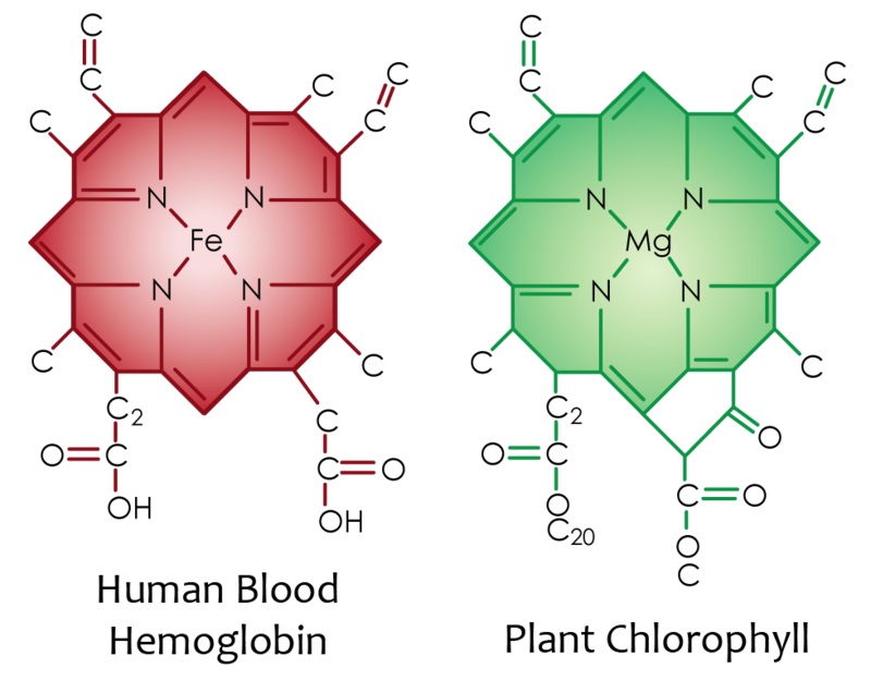
To explain this to those who lack the scientific background in Crop/Human Bio-Chemistry, he would explain that these poor crops lead to the body up taking excess Nitrogen, in the form of Nitrite, which is a chief player in lowered Oxygen status within the body. He would explain how plants yield Haemoglobin supportive substances (especially green leafy) such as Chlorophyll which is Magnesium at it’s centre, surrounded by a shell which is virtually chemically identical to Haemoglobin, so the body may use this readily to create this Heme with Magnesium being replaced by Iron (simple body alchemy), the Magnesium may then be used in ATP production (pertinent to this dynamic). The Haemoglobin is that which allows Oxygen to bind to it within the Bloodstream, and carry it into tissue, conversely, Methaemoglobin is it’s Oxygen uptake antagonised state (Fe3+ Ferric once Oxidised becomes Methemoglobin Vs Fe2+ Ferrous where normal Haemoglobin) the Iron is oxidised it becomes compromised.
Cursory research of the scientific literature, shows that issues with Methaemoglobin, induces Oxygen desaturation, Cyanosis, and is often Nitrogen driven (this is why Nitrogen levels in drinking water are controlled, though many companies sell mineral/spring water with levels way above that which is healthful).
The blood of those with good Oxygen saturation may be bright red, those with high Methaemoglobin may show blood which is brown with blueness. Showing this state in action. The blueness aspect is how humans may externally show that there is imbalance, blue lips, around the eyes blueness and brown circles.
Sadly, although they existed and were active research (and application of findings) Scientists at the very same timeframes, to my knowledge Reams and Budwig never communicated, though it seems certain Budwig was aware of Reams and talked of how “those who advocate Olive Oil are very unhelpful”, talking of this dynamic. Reams advocated light dinners of Salad with Olive Oil (showing his inferior understanding of lipid dynamics) to allow the digestion rest and ready itself for the following day, no protein rich foods later as the digestion of these uses more Oxygen, leaving potentially less as we sleep if the digestion is unfinished, circadian rhythm shows that the adrenals long for oxygen to repair and replenish from 11pm-1am giving credence to this thinking.
Conversely, Budwig lacked the deeper understanding of plant matter that Reams had, how high nutrient density played such a crucial role in that very same function of optimal Oxygen conductance and uptake in the human body. Both geniuses of which I am hardly qualified to talk of, let alone critique, so please know it is just a longing for the fuller picture to emerge from science which draws out these words, not negativity to either giant of nutritional healing.
So, it is easy to see, the inherent pitfalls of many modern techniques for returning to well being, given the excessive saturation of poor crops on the vegetable side, lacking nutrient density, being farmed for high yield, early, to allow cheap Supermarket produce which we all see is often rotting by the time you get them home. Likewise meats grown on these poor crops/lands, which pushes their tissue Oxygen saturations lower and heightens their Nitrate status for the consumer. On the lipid side, Flaxoil in most shops are not vitally fresh, seeds are old and pre-ground when Budwig said they should be fresh ground, soo much of our produce is coated in Oxygen antagonistic lipids.
Free Electrons. Budwig may have been on to something???https://www.mriquestions.com/deoxy-hb-v-met-hb.html
When it is discussed “who would your dream dinner party guests be” Reams and Budwig are always two of mine, if only they could have been locked in a room together for a few days, a broader pioneering truth would surely have emerged.
Acidosis is a complex metabolic state with a range of etiologies (Planche and Krishna 2006; Taylor et al. 2012). Within malaria, acidosis is caused by a combination of several factors. The malaria parasite produces Plasmodiumlactate dehydrogenase (pLDH), which creates lactic acid leading to decreased pH. Respiratory distress is a common feature of severe malaria and, through sequestration, somnolence, and/or brain swelling, direct central suppression of the respiratory centers leads to irregular breathing patterns in the setting of acidosis, which may contribute to the pH imbalance. Supportive therapy to protect the airway and more aggressively rebalance the pH may decrease mortality (Cheng and Yansouni 2013).