FDA-approved artificial sweeteners and sport supplements were found to be toxic to digestive gut microbes, according to a new paper published in Molecules by researchers at Ben-Gurion University of the Negev (BGU) in Israel and Nanyang Technological University in Singapore.
The collaborative study indicated relative toxicity of six artificial sweeteners (aspartame, sucralose, saccharine, neotame, advantame, and acesulfame potassium-k) and 10 sport supplements containing these artificial sweeteners. The bacteria found in the digestive system became toxic when exposed to concentrations of only one mg./ml. of the artificial sweeteners.
“We modified bioluminescent E. coli bacteria, which luminesce when they detect toxicants and act as a sensing model representative of the complex microbial system,” says Prof. Ariel Kushmaro, John A. Ungar Chair in Biotechnology in the Avram and Stella Goldstein-Goren Department of Biotechnology Engineering, and member of the Ilse Katz Institute for Nanoscale Science and Technology and the National Institute for Biotechnology in the Negev. “This is further evidence that consumption of artificial sweeteners adversely affects gut microbial activity which can cause a wide range of health issues.”
Artificial sweeteners are used in countless food products and soft drinks with reduced sugar content. Many people consume this added ingredient without their knowledge. Moreover, artificial sweeteners have been identified as emerging environmental pollutants, and can be found in drinking and surface water, and groundwater aquifers.
“The results of this study might help in understanding the relative toxicity of artificial sweeteners and the potential of negative effects on the gut microbial community as well as the environment.
Furthermore, the tested bioluminescent bacterial panel can potentially be used for detecting artificial sweeteners in the environment,” says Prof. Kushmaro.
More information: Dorin Harpaz et al, Measuring Artificial Sweeteners Toxicity Using a Bioluminescent Bacterial Panel, Molecules (2018). DOI: 10.3390/molecules23102454
ChooseLife Reflects : When our daughter (after her extreme premature arrival) was in hospital, it was relayed that they routinely gave Sucralose after taking bloods, as an Australian study had shown benefits to the babies emotional wellbeing afterwards.
This shocked me as I am strongly against their use (personally), so I relayed that I would rather they did not.
The day following the Consultant, several Doctors and Nurses came to see us to relay that they were not not happy with my/our refusal. My response was to break the compound down chemically and relay that it is not found in nature, so I did not want it for our daughter, that it was an adjunct to a treatment which did not have any effect on the efficacy of the procedure (therefore optional). The Consultant acknowledged that I had researched things and this was therefore fine.
It is my belief that staff were drawn to it’s use, as they noted smiles and the like from the babies, ignoring the potential biochemical issues which may also ensue. The nurses felt better about stabbing these little babies to withdraw blood, as they left with babies appearing happier, with less tears and crying, ignoring the wider picture.
Glyphosate is among the most widely used agricultural chemicals in the world, and the companies that make it describe it as safer and more environmentally-friendly than other herbicides.
But a group of researchers in Texas now report that the weed killer, sold as Roundup, could harm honeybees indirectly by disrupting the bacteria in their guts (Proc. Natl. Acad. Sci. USA 2018, DOI: 10.1073/pnas.1803880115). The scientists think the findings could help explain the decline in honeybees observed over the past decade or so.
The team, led by Nancy Moran, an expert in bee biology at the University of Texas, Austin, treated hundreds of worker bees with glyphosate at concentrations they might encounter while foraging near agricultural fields, reintroduced them into their hive, and then analyzed the gut bacteria of treated and untreated bees in the hive. After three days, the researchers found that the abundance of some of the eight predominant species fell in treated bees compared with untreated ones, suggesting that exposure to glyphosate changed the composition of the bees’ guts. When challenged with a common bee pathogen, the worker bees exposed to glyphosate died at higher rates than unexposed bees, suggesting that the bacterial shake-up made the insects more vulnerable.
The conclusions match previous reports demonstrating that honeybees with compromised gut flora are malnourished and susceptible to infection. Those findings, along with data showing that glyphosate can affect soil bacteria and accumulate in bee honey and hives, indicate that researchers should explore whether possible off-target effects of the popular herbicide have played a role in honey bee decline, Moran says.
About 10 years ago, there was a noted die-off of honeybees, later termed colony collapse disorder. Mortality rates have continued to be high, Moran says, but the die-offs haven’t been as sudden. Researchers have been looking at the role of pesticides and fungicides in the decline, as well as infection. But glyphosate hasn’t really been part of those earlier conversations, says Fred Gould, an entomologist and plant pathologist at North Carolina State University. The field, he says, is “getting better at looking for subtle off-target effects,” such as those described in the new study.
As an herbicide, glyphosate works by blocking an enzyme called EPSP synthase in plants and microbes needed to create aromatic amino acids like phenylalanine and tryptophan. Glyphosate doesn’t kill microbes, but it keeps them from growing, and most bacteria found in bee guts carry the gene for the enzyme.
The environmental impact and health effects of glyphosate form a contentious issue, Gould says, so whole hive studies could shed light on the herbicide’s effects on a critical species for agriculture.
These studies also could help guide how to avoid possible negative effects of glyphosate on honeybees. The likely outcome will be some sort of best practices on when and where to spray glyphosate, says Juliana Rangel, an entomologist at Texas A&M University. Pollination via honeybees is an approximately $15 billion per year industry, she says, and “a lot of times, these tragedies can be avoided with a better understanding of the system.”
Diets high in carbohydrates reduce body weight and body fat and improve insulin function in overweight individuals, according to a new study published in Nutrients.
In the 16-week randomized clinical trial, researchers with the Physicians Committee for Responsible Medicine placed participants in either a plant-based, high-carbohydrate, low-fat diet group or asked them to maintain their current diet. The plant-based diet group avoided all animal products and added oils and limited fat intake to 20-30 grams per day. There were no limits on calories or carbohydrate intake. The control group maintained their current diets, which included meat and dairy products. Neither group altered their exercise routines.
Total carbohydrate intake did not change in the control group, but increased significantly in the plant-based diet group, both as absolute intake and as a percentage of total calories. Participants focused on whole, complex carbohydrates from fruits, vegetables, whole grains, and legumes.
At the end of the trial, body mass index, body weight, fat mass, visceral fat volume, and insulin resistance decreased significantly in the plant-based diet group. There were no significant changes in the control group.
“Fad diets often lead people to fear carbohydrates. But the research continues to show that healthy carbohydrates–from fruits, vegetables, beans, and whole grains–are the healthiest fuel for our bodies,” says lead study author Hana Kahleova, M.D., Ph.D., director of clinical research for the Physicians Committee for Responsible Medicine.
The study’s results support previous research finding that a plant-based, high-carbohydrate diet can help with weight regulation and body composition and reduce the risk for type 2 diabetes. Another recent study published in The Lancet found that people who consume animal-based, low-carbohydrate diets have a shorter life expectancy, compared with those who consume more carbohydrates and/or more plant-based sources of protein or fat.
Complex carbohydrates are naturally rich in fiber–a nutrient found in plant foods that adds bulk to the diet without adding extra calories. Previous studies have shown that high-fiber diets are effective for weight loss and can help reduce the risk for type 2 diabetes, heart disease, and certain types of cancer.
The study has important implications, because more than 7 in 10 U.S. adults are considered overweight or obese. Approximately 30 million Americans have diabetes, while prediabetes affects 84 million more.
Summary : Body organs such as the intestine and ovaries undergo structural changes in response to dietary nutrients that can have lasting impacts on metabolism, as well as cancer susceptibility.
Body organs such as the intestine and ovaries undergo structural changes in response to dietary nutrients that can have lasting impacts on metabolism, as well as cancer susceptibility, according to Carnegie’s Rebecca Obniski, Matthew Sieber, and Allan Spradling.
Their work, published by Developmental Cell, used fruit flies, which are currently the most-sensitive experimental system for such detecting diet-induced cellular changes that are likely to be similar in mammals.
There are three major types of cells in fruit fly (and mammalian) intestines: Stem cells, hormone-producing cells, and nutrient-handling cells. Think of the stem cells as blanks, which are eventually programmed to become either hormone-producing or nutrient-handling cells. The authors discovered that this programming can be influenced by dietary nutrients, and that young animals are particularly sensitive to these changes.
Obniski, the lead author, and her colleagues found that changes in dietary cholesterol particularly alter the cellular programming driving the production of new specialized cells from stem cells.
The effect of cholesterol is to promote the programming of more new, “blank” cells into hormone-producing cells rather than nutrient-handling cells. Conversely, decreasing dietary cholesterol results in more nutrient-absorbing cells and fewer hormone-producing cells.
Moreover, the researchers were able to identify the detailed molecular mechanism by which cholesterol causes these changes in cell fates, and to show that it is closely related to the way human intestinal cells regulate cholesterol production.
What does this mean?
It shows that low nutrient availability, especially early in life, such as the low-cholesterol diet for the fruit flies, triggers changes in intestinal structure and metabolism that have long-term effects. These changes persist for quite a while even if the diet changes, which can increase the risk of metabolic health problems down the road.
“Children born to malnourished mothers often struggle with obesity later in life and our findings could explain the physiology of why that happens,” Obniski explained.
She and her colleagues say that further understanding the how nutrient availability affects intestinal function could help researchers find ways to use diet to mitigate aging and disease in adults.
For example, the biochemical signaling pathways that were shown to underpin this developmental metabolic programming, explain why a high-fat diet can promote the formation of certain types of intestinal cancer.
“The intestines are the organs most-directly responsible for balancing an organism’s long- and short-term needs with its nutritional environment,” Spradling said. “The power of the fruit fly as a model system allows one of the complex mechanisms that balance these needs with diet to be understood at a mechanistic level, something that is very difficult to do directly in mammalian systems or in human patients.”
Discover silica’s anti-aging and heavy metal detox properties
(NaturalNews) The mineral silica (Si) is getting more notice for its important functions. Sometimes called the “beauty mineral” because it improves skin elasticity and hair and nail growth, a few other more important aspects have been explored lately.
Silica helps ensure collagen elasticity of all connecting tissues in the body, including tendons and cartilage. This reduces aches and pains and maintains your body’s flexibility. It has also been determined lately that high levels of blood serum silica keep arterial plaque from building and clogging blood vessels.
The main culprit for that plaque has recently shifted from cholesterol buildup to calcification from calcium in the blood that is not absorbed as bone matter. It has been known that silica is an important part of building bone matter. Without it, calcium goes elsewhere to potentially calcify in the soft tissue of inner artery walls and the heart.
Silica is vital for keeping strong bones and a healthy cardiovascular system. This qualifies silica as an essential anti-aging mineral that is much more than skin deep.
Silica for detoxing heavy metals – especially aluminum
Even PubMed acknowledges silicic acid as an antidote for aluminum poisoning and silica as a detox agent for heavy metals. Both brain tissue calcification and aluminum toxicity are linked to Alzheimer’s disease.
Now it’s been discovered that silica can be supplemented to help prevent yet another aspect of aging – dementia. Obviously it can also be used as an adjunct with any other Alzheimer’s treatment, such as pure cold pressed coconut oil (https://www.naturalnews.com/030919_coconut_oil_Alzheimers.html).
Aluminum (Al) is passed out through the urine when one supplements silica. It seems there’s little danger of taking too much, as long as adequate water is consumed and vitamin B1 and potassium levels are maintained.
There is no shortage of aluminum toxicity in our environment. It’s in cookware, beverage containers, foil, second hand cigarette smoke, cosmetics, sunscreen, chemtrails, and more. For sure, it is in all vaccines. Injecting aluminum bypasses the normal route of eliminating it.
Aluminum accumulates in tissue that doesn’t have a rapid cellular turnover. The slow turnover tissues are contained in bone matter, the heart and the brain. The brain and its associated nervous system is where diseases such as Parkinson’s, MS, chronic fatigue and other neurological or auto-immune diseases manifest.
Dr. Chris Exley, PhD, has dedicated almost two decades of his scientific life to researching aluminum toxicity. He calls the period of time from the early 20th Century to now the “Age of Aluminum.” Before then, aluminum remained in the ground and hadn’t yet been mined. Exley claims mining aluminum and using it in so many ways corresponds to the marked increase of neurological diseases.
He recommended a couple of pricey mineral waters high in silica that better health food stores carry (Video source below, Exley lecture). He claimed positive results occurred with vaccine injured children drinking these waters, but there are other good less pricey silica sources as well.
Silica sources
Horsetail is an excellent and inexpensive herbal source of silica. Horsetail has been around for centuries, and mostly forgotten until recent research attention uncovered more attributes. Until then, horsetail was used mostly as a diuretic or treatment for kidney stones.
Anything for which silica is useful will benefit from horsetail. And there are supplements that are formulated for silica intake that you may want to look into. Foods that help keep your silica levels high are: Unrefined whole grains such as rye, barley, oats, and wheat. Alfalfa sprouts nuts, and other seeds will boost your silica levels.
About the author:
Paul Fassa is dedicated to warning others about the current corruption of food and medicine and guiding them towards direction for better health with no restrictions on health freedom.
(NaturalNews) How much aluminum is in your drinking water? It’s hard to tell, but in a 15-year study on French elderly men and women, regular consumption of tap water was associated with aluminum toxicity and increased prevalence of dementia. How might the accumulation of aluminum from just tap water alone affect your cognitive ability as you age?
In the 15 years of study, researchers found out that dietary aluminum from water sources could be a risk factor for developing dementia. They also found a helpful solution. They found that the mineral silica could help decrease the risk of aluminum-induced dementia in elderly patients.
A 2009 report from the American Journal of Epidemiology details this 15-year study from France. The study followed the lives of 1,925 elderly patients between the years 1988 and 2003. All of the elderly men and women were selected from 91 different municipalities with different water sources in southern France. Their aluminum intake levels were studied, from water and other sources, as the researchers investigated the metal’s role in bringing about dementia throughout the 15-year period. During that time, the researchers also investigated the impact of silica in reducing participants’ risk of dementia.
Aluminum from drinking water increased dementia in 15-year study
Of the nearly 2,000 elderly studied, none showed any signs of dementia beginning in 1988.
The researchers ruled out environmental aluminum intake factors during the study and focused solely on aluminum intake from water. The participants’ daily consumption of tap and bottled water was recorded. Reliable water assessment data was analyzed. In the study, aluminum consumption greater than or equal to 0.1 mg per day from drinking water was correlated with declining cognitive ability. Over the years, the accumulation of aluminum inflicted negative mental effects, welcoming dementia in what was once a mentally healthy group of elderly adults. Using the scientific Cox model, the researchers found that highest exposure to aluminum may be a risk factor for full-blown dementia.
Hoping to the help the participants, the researchers looked further and assessed silicaintake for its ability to reduce dementia.
What they discovered was that when participants increased their silica intake by 10 mg/day, incidences of dementia subsided. The change was drastic.
This discovery coincides with the expert analysis of Dr. Chris Exley, PhD.
Doctor recommends silica to help those affected by aluminum from vaccines
At a January 2011 vaccine safety conference in Jamaica, Dr. Exley talked about ways to reduce the toxic effects of aluminum in the body. He talked about the importance of silica and mentioned two brands of silica-enhanced water, Volvic and Spritzer. After conducting several urine tests, Exley and his team of researchers proved that high-silica mineral waters help remove aluminum from the body. In a presentation involving victims of Gardasil’s adverse reactions, Exley showed how silica improved the damages caused by aluminum-laced vaccines. Based off 20 years of study, Dr. Exley recommends drinking a liter of silica-rich water daily to reduce the burden of accumulated aluminum in the body.
Sources of silica
Thankfully, silica mineral water isn’t the only place to obtain this important mineral.
According to the 1993 book Silica: The Forgotten Nutrient, by Klaus Kaufmann, silica can be found primarily in the following foods (Silica content is measured in mg.):
Oats: 595
Millet: 500
Barley: 233
Potatoes: 200
Whole wheat grain: 158
Jersusalem artichoke: 36
Red beets: 21
Corn: 19
Asparagus: 18
Rye: 17
Horsetail – top source for silica
One of the sources of silica not mentioned above is horsetail. Respected as a strong diuretic and astringent herb, horsetail is a fern-like, nonflowering weed that is loaded with silica. The tall, hollow stems resemble asparagus and often grow in bunches. When the plant begins to dry, silica crystals form in the stems and appear to look like feathery tails. This herb can be purchased as a dry powder and can be used raw to make tinctures. The silica content of horsetail is unmatched and is famous for helping people grow healthy hair, nails and skin.
With its high silica content, horsetail may also be good for removing aluminum from the body and warding off dementia.
Kosinski : “If I know 70 of your likes on Facebook, that’s enough to outdo what your friends know about you. If I know 150 likes that you clicked on Facebook, I know you better than your parents know you.” And at 300 likes, Kosinski intimated, the algorithm knows you better than you know yourself.
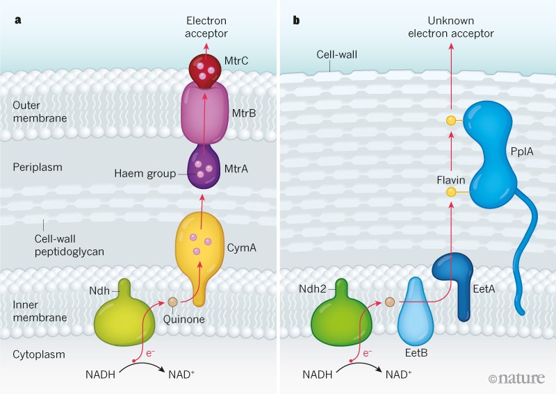
Some bacteria make energy in a process that is accompanied by transfer of electrons to a mineral. A previously unknown electron-transfer pathway now reveals an energy-generation system used by bacteria in the human gut.
The ability of certain bacteria to transfer electrons has been exploited for a variety of energy-generating applications, such as microbial fuel cells1, because the flow of charge carried by electrons underlies the process that generates electricity. It was thought that the capacity to achieve substantial levels of electron transfer occurred only in a specialized subset of bacteria. These microbes make energy by a mechanism that requires minerals for the electron-transfer process that accompanies energy generation2. Writing in Nature, Light et al.3 report the discovery of an electron-transfer pathway in gut bacteria, and reveal that components of this pathway are present in diverse microbial species.
The molecule ATP provides the fundamental energy ‘currency’ for most cells, and is mainly produced by two mechanisms: fermentation, an anaerobic process in which ATP is generated from a limited repertoire of carbon sources, and respiration, a process that provides a high yield of ATP from a wide array of carbon sources and requires a compound that can accept electrons. In multicellular organisms, respiration involves electron transfer along an electron-transport chain that culminates in electrons being transferred to oxygen4.
By contrast, microbes can use a number of alternatives to oxygen as electron acceptors that enable respiration in anaerobic environments lacking fermentable energy sources2,5. For example, the bacteria Shewanella oneidensis and Geobacter metallireducens reside in mineral-rich environments, and these highly studied microbes have an anaerobic respiration process that uses minerals, such as iron(iii) oxide (Fe2O3), as respiratory electron acceptors2. However, because insoluble mineral deposits cannot be transported into the cell, mineral-respiring bacteria use a mechanism2 called extracellular electron transfer (EET), in which electrons are transferred to the exterior of the cell. In the case of these bacteria, this process involves electron transfer from an NADH molecule to components that include a quinone molecule in the lipid membrane and a series of proteins containing haem groups that provide a path for electron transfer. The loss of an electron converts NADH to NAD+, which is used in the energy-generation process.
The food-borne bacterial pathogen Listeria monocytogenes sometimes has a host-associated part of its life cycle. This bacterium can infect humans, and can proliferate in nutrient-rich environments that enable the use of fermentation as a metabolic strategy6. However, although L. monocytogenes has a life cycle in which neither minerals nor respiration is crucial for survival, Light et al. report that, when L. monocytogenes was placed in an electrochemical chamber in which an electrode can trap electrons, an electric current was generated, suggesting that this type of bacterium has the capacity for EET. This report now clarifies evidence presented decades ago7, indicating that this bacterium can change extracellular iron in the Fe3+ form to the Fe2+ form, an alteration that might indicate electron transport out of the cell.
Using a combination of genetic and biochemical approaches, Light et al., true to the name, shed light on the molecular basis of this newly discovered form of EET. They identified the proteins Ndh2, EetB, EetA and PplA as being key components of this process. They show that the initial electron-transfer steps of EET in L. monocytogenes resemble those already known in mineral-respiring specialists. For example, electron transfer from the cell cytoplasm to a quinone molecule in the lipid membrane is similar to the steps of a conventional electron-transport chain. However, beyond this point, the mechanisms become more distinct. L. monocytogenes is a Gram-positive bacterium, which means that it has a single lipid membrane and a thick cell wall. By contrast, S. oneidensis and G. metallireducens are Gram-negative bacteria, which have two lipid membranes separated by a region called the periplasm that contains cell-wall material. In these bacteria, tens of haem molecules bound to three types of protein establish a path for electrons to move across the periplasm and the outer lipid membrane8. By contrast, in L. monocytogenes, a single protein called PplA that contains two flavin molecules suffices to enable electrons to exit the membrane to reach the cell’s exterior (Fig. 1).
Figure 1 | Bacterial electron-transfer pathways.a, In the Gram-negative bacterium Shewanella oneidensis, the mechanism that generates energy from the molecule NAD+ is accompanied by a process in which an electron (e−) from the molecule NADH is transferred outside the cell to a mineral such as iron(iii) oxide that acts as an electron acceptor. In this process, known as extracellular electron transfer (EET), the electron-transfer path (red arrow) occurs across two lipid membranes and across the periplasm region, which contains cell-wall material that includes the sugar peptidoglycan. The electron-transfer route towards the cell exterior after crossing the protein Ndh includes a quinone molecule, haem groups associated with the proteins CymA, MtrA and MtrC, and transfer through the protein MtrB.b, Light et al.3 report a previously unknown EET mechanism in Listeria monocytogenes, a Gram-positive bacterium, which has only a single membrane. The authors identified components of this EET system, including the proteins Ndh2, EetB, EetA and PplA (which is associated with two flavin molecules). This newly identified EET process might occur in diverse bacteria, including those in the human gut. The electron acceptor for the pathway is unknown.
Light and colleagues analysed the distribution of the genes for this newly identified EET pathway in the genomes of different bacterial species, and provide evidence of EET activity in species other than L. monocytogenes using an electrochemical chamber. They reveal that EET activity occurs in an environmentally and evolutionarily diverse subset of Gram-positive bacteria, most notably in certain bacteria found in the human gut, such as those of the genus Lactobacillus.
This observation is intriguing because EET usually provides energy in anaerobic conditions, and growth strategies for such conditions can be important for microbial proliferation in the mammalian gut9. Indeed, Light et al. found that genes encoding components of the EET system they identified are required for L. monocytogenes to grow in anaerobic conditions. Moreover, when the authors monitored the ability of L. monocytogenes strains to colonize the mouse gut, the strains deficient in components of this EET system were at a competitive disadvantage, suggesting that EET has a key role in bacterial survival in this context. Investigating the role of EET in host–microbe interactions could offer an exciting direction for future research.
A central question raised by these findings is why EET might have evolved outside the context of mineral-respiring specialists. The bacterial environment may provide a clue. When microbes such as L. monocytogenes live in a host gut, they are immersed in nutrients, including flavin molecules, and Light et al. show that the presence of flavins potently enhances EET activity. The electron-transfer apparatus is simpler in Gram-positive bacteria than in Gram-negative bacteria. It stands to reason that an abundance of environmental flavins might produce a scenario in which evolution favours the minimal investment in protein infrastructure needed to enable EET in certain Gram-positive bacteria. EET might be used by certain mineral-respiring bacteria because it is crucial for their survival, whereas L. monocytogenes might use EET because it provides an opportunity to easily generate energy in certain environments.
The electron acceptor used by L. monocytogenes for EET is unknown. The bacterium might encounter conditions in which minerals represent an attractive electron acceptor, but it seems more probable that the highly reactive flavins in this pathway aid electron transfer to compounds such as organic soil components, disulfide groups on proteins or even other microbes10,11. If this is the case, in contrast to EET associated with specialized mineral respiration, the EET in L. monocytogenes might provide a more flexible mechanism for moving electrons to a variety of environmental acceptors.
It is a shock to the system to consider that microbes might be living highly charged lives in our gut. Light and colleagues’ work provides a foundation for future investigation regarding such microbial existence. Furthermore, the characterization of this previously unknown EET mechanism might create opportunities for the design of bacteria-based energy-generating technologies.
Summary : Pollinating insects are endangered globally, with a particularly steep decline over the last 40 years. An extensive 3-year study has found that organic farming methods can contribute to halting the pollinator decline. This beneficial effect is due to both the absence of insecticides and a higher provision of flower resources.
Organic farming is known to promote pollinator diversity in crop fields. However, it has also been suggested that organic fields might simply attract pollinators from other habitats in the landscape, and therefore not sustain their populations in the long run.
The 3-year field experiment, conducted by researchers from the Centre for Environmental and Climate Research at Lund University, found that the number of bumblebee species in organic farms was higher and more stable over time and space than in conventional farms.
“This is the first large-scale study over the course of several years to show that organic farming has a consistent, stabilizing effect on pollinator diversity ,” says Romain Carrié, a postdoctoral researcher at CEC.
Romain and his colleagues sampled bumblebees, butterflies and flowering plants throughout the growing season in 10 organic and 9 conventional farms in Scania, Sweden. Their study showed that, depending on the type of crop, the stabilizing effect was either due to a more stable provision of flowers or the absence of pesticides.
“An interesting result of our study is the fact that stable and abundant flower resources stabilizes pollinator communities, even in conventional farms where insecticides are used,” explains Romain Carrié.
“This is strongly suggesting that both flower-enhancing management options and a reduced use of insecticides can help reverse pollinator declines,” Romain Carrié concludes.
Renal diets for advanced chronic kidney disease (CKD) are structured to achieve a lower protein, phosphate and sodium intake, while supplying adequate energy. The aim of this nutritional intervention is to prevent or correct signs, symptoms and complications of renal insufficiency, delaying the start of dialysis and preserving nutritional status. This paper focuses on three additional aspects of renal diets that can play an important role in the management of CKD patients: the vitamin K1 and fiber content, and the alkalizing potential. We examined the energy and nutrients composition of four types of renal diets according to their protein content: normal diet (ND, 0.8 g protein/kg body weight (bw)), low protein diet (LPD, 0.6 g protein/kg bw), vegan diet (VD, 0.7 g protein/kg bw), very low protein diet (VLPD, 0.3 g protein/kg bw). Fiber content is much higher in the VD and in the VLPD than in the ND or LPD. Vitamin K1 content seems to follow the same trend, but vitaminK2 content, which could not be investigated, might have a different pattern. The net endogenous acid production (NEAP) value decreases from the ND and LPD to the vegetarian diets, namely VD and VLPD; the same finding occurred for the potential renal acid load (PRAL). In conclusion, renal diets may provide additional benefits, and this is the case of vegetarian diets. Namely, VD and VLPD also provide high amounts of fibers and Vitamin K1, with a very low acid load. These features may have favorable effects on Vitamin K1 status, intestinal microbiota and acid-base balance. Hence, we can speculate as to the potential beneficial effects on vascular calcification and bone disease, on protein metabolism, on colonic environment and circulating levels of microbial-derived uremic toxins. In the case of vegetarian diets, attention must be paid to serum potassium levels.
KEYWORDS:
CKD; PRAL; Renal diets; Vitamin K1; fiber; gut microbiota; low protein diet, renal nutrition, metabolic acidosis; uremic toxins
TUESDAY, Sept. 4, 2018 (American Heart Association) — A varied, quality diet could help prevent hospitalizations and even death among patients with heart failure, a new study suggests.
Researchers investigating nutritional deficiencies found that people with heart failure who lack seven or more micronutrients had nearly double the risk of dying or being hospitalized than those who didn’t have any or only a few deficiencies. The University of Kentucky-led study was published Sept. 4 in the Journal of the American Heart Association.
“This establishes the importance of nutrition and why it really has to become a higher priority when it comes to treating heart failure,” said lead author Terry Lennie, senior associate dean at the University of Kentucky’s College of Nursing. “Nutritional deficiencies really can put patients at risk, more so than I think we understood or appreciated before.”
The study examined data from 246 patients recruited from three heart failure clinics in Georgia, Indiana and Kentucky. Patients kept detailed diaries of everything they ate and drank for four consecutive days.
Researchers assessed the intake of 17 micronutrients — 11 vitamins and six minerals — from the food diaries. They also kept tabs on patients every month for the following year.
The study found that 44 percent of patients with deficiencies in seven or more micronutrients were hospitalized or died within the year, compared to 25 percent of patients who had no deficiencies or only a few.
Calcium was the most commonly deficient micronutrient in patients’ diets, followed by magnesium, vitamins D and E, zinc and vitamin C.
One reason for the lack of these micronutrients could be “diet monotony,” or the tendency to eat the same foods every day instead of incorporating variety into meals. The study found many patients consumed the same foods for multiple meals across all four days of the food diary. Older adults are more vulnerable to this habit “due to a decreased drive to consume varied foods,” the study said. The average age of patients was 61.
A majority of the participants were overweight or obese, dispelling the notion of a link between a person’s weight and nutritional deficiencies.
“When we see individuals who are overweight, people tend to think they’re well-nourished, and that we only have to worry about people who are underweight as far as nutrition goes,” Lennie said. “But we found no relationship between patients’ body mass index and whether or not they had nutritional deficiencies.”
Dr. Frank Hu said the use of four-day food diaries did a good job capturing patient dietary patterns. But Hu, chairman of the nutrition department at Harvard University’s T.H. Chan School of Public Health, said he would have liked to have seen a much larger study size.
Hu, a professor of nutrition and epidemiology who was not involved in the research, said the findings demonstrate the role that well-rounded, varied diets can play in keeping heart failure patients alive. He noted the study did not address whether any single nutrient played a more important role than others. It instead looked at overall dietary health.
“It’s very important to pay attention to both nutrition quantity and quality. When we talk about nutrition quality, we’re not talking about just popping a vitamin or mineral supplement,” he said.
“Micronutrients come mostly from plant-based foods, such as fruits and vegetables, whole grains, nuts and legumes,” Hu said, adding that including some animal foods such as moderate amounts of fish and dairy products is also helpful in achieving adequate micronutrient intakes.
“It’s more important to pay attention to the quality of the foods when we try to make sure patients eat a balanced, nutritional meal,” he said.
Last Updated:
Copyright is owned or held by the American Heart Association, Inc., and all rights are reserved. If you have questions or comments about this story, please email editor@heart.org.