
Functional Role of Dietary Intervention to Improve the Outcome of COVID-19: A Hypothesis of Work
Authors : Giovanni Messina (1), Rita Polito (1), Vincenzo Monda (2), Luigi Cipolloni (1), Nunzio Di Nunno (3), Giulio Di Mizio (4), Paolo Murabito (5), Marco Carotenuto (6), Antonietta Messina (2), Daniela Pisanelli (1), Anna Valenzano (1), Giuseppe Cibelli (1), Alessia Scarinci (7), Marcellino Monda (2), Francesco Sessa (1)
- Department of Clinical and Experimental Medicine, University of Foggia, Italy
2. Department of Experimental Medicine, Section of Human Physiology and Unit of Dietetics and Sports Medicine, Università degli Studi della Campania Naples, Italy
3. Department of History, Society and Studies on Humanity, University of Salento, Lecce Italy
4. Department of Law, Forensic Medicine, Magna Graecia University of Catanzaro, Italy
5. Department of General Surgery and Medical-Surgical Specialties, University of Catania, Italy
6. Department of Mental Health, Physical and Preventive Medicine, Clinic of Child and Adolescent Neuropsychiatry, Università degli Studi della Campania Italy
7. Department of Education Sciences, Psychology and Communication, University of Bari, Italy
Abstract
Background: On the 31 December 2019, the World Health Organization (WHO) was informed of a cluster of cases of pneumonia of unknown origin detected in Wuhan City, Hubei Province, China.
The infection spread first in China and then in the rest of the world, and on the 11th of March, the WHO declared that COVID-19 was a pandemic. Taking into consideration the mortality rate of COVID-19, about 5–7%, and the percentage of positive patients admitted to intensive care units being 9–11%, it should be mandatory to consider and take all necessary measures to contain the COVID-19 infection.
Moreover, given the recent evidence in different hospitals suggesting IL-6 and TNF-α inhibitor drugs as a possible therapy for COVID-19, we aimed to highlight that a dietary intervention could be useful to prevent the infection and/or to ameliorate the outcomes during therapy. Considering that the COVID-19 infection can generate a mild or highly acute respiratory syndrome with a consequent release of pro-inflammatory cytokines, including IL-6 and TNF-α, a dietary regimen modification in order to improve the levels of adiponectin could be very useful both to prevent the infection and to take care of patients, improving their outcomes.
1. Background
On the 31 December 2019, the World Health Organization (WHO) was informed of a cluster of cases of pneumonia of unknown origin detected in Wuhan City, Hubei Province, China. About one month later (on 8 January 2020), the Chinese authorities declared the identification of a new type of coronavirus, informing the WHO a few days later that the outbreak was associated with exposure in a seafood market in Wuhan City.
The infection spread firstly in China and then in the rest of the world, and on the 11th of March, the WHO declared that COVID-19 was a pandemic.Coronaviruses (CoVs) belong to the subfamily Orthocoronavirinae in the family of Coronaviridae in the order Nidovirales, and this subfamily includes α-coronavirus, β-coronavirus, γ-coronavirus, and delta-coronavirus [1].
Coronaviruses primarily cause enzootic infections in birds and mammals and, in the last few decades, have shown to be capable of infecting humans as well [2]. In human infections with highly virulent respiratory viruses—such as avian influenza H5N1, H7N9, Severe Acute Respiratory Syndrome (SARS) coronavirus, and Coronavirus Disease-19 (COVID-19)—immunopathogenesis caused by the overproduction of pro-inflammatory cytokines may play an essential role in disease progression and mortality [3].
Several recent studies have reported that COVID-19 caused the destruction of the pulmonary parenchyma, including interstitial inflammation and extensive consolidation, similarly to the previously reported coronavirus infection [4,5]. During coronavirus infection, it was observed that the lungs increased in weight, with a mild pleural effusion of clear serous fluid, named pulmonary edema, and extensive consolidation [6,7]. In some areas, there was interstitial thickening, with mild-to-moderate fibrosis, but a disproportionately sparse infiltrate of inflammatory cells (mainly histiocytes, including multinucleated forms, and lymphocytes) [8]. A dilatation of the airspaces was observed, as was focal honeycombing fibrosis. An intra-alveolar organization of exudates was described, and the formation of granulation tissues in the small airways and airspaces was reported. These lesions were typically located in the sub-pleural region, and the cellular component mainly consisted of histiocytes, as reported in a previous paper [9]. Xu et al. described in their case report the pathological findings of COVID-19 associated with acute respiratory distress syndrome. At the X-ray investigation, they detected a rapid progression of bilateral pneumonia.
The biopsy samples were taken from the lung; the histological examination showed bilateral diffuse alveolar damage with cellular fibromyxoid exudates [6].Considering that the mortality rate of COVID-19, about 5–7% [10], and the percentage of positive patients admitted to intensive care units being 9–11% [11], it should be mandatory to consider and take all necessary measures intended to contain the viral infection.
A recent study analyzed the data of 150 COVID-19 patients, with the aim of defining the clinical predictors of mortality. The results obtained from this study suggest that COVID-19 mortality might be due to virus-activated “cytokine storm syndrome”, considering that the plasma levels of IL-6 were higher in deceased patients compared to in discharged subjects [12].Considering that a detailed study has not been performed on the immunological response to COVID-19, the only way to discuss this thematic is to refer to previous knowledge about SARS-CoV and MERS-CoV. The first response is obtained through pattern recognition receptors (PRRs) including C-type lectin-like receptors, Toll-like receptors (TLR), NOD-like receptors (NLR), and RIG-I-like receptors (RLR). Moreover, several inflammatory factors are expressed such as IL-6 and TNF-α; moreover, the synthesis of type I interferons (IFNs) is activated, and these exert their actions against virus diffusion, accelerating macrophage phagocytosis [13] (Figure 1).

In the light of these considerations and the recent evidence in different hospitals suggesting IL-6 and TNF-α inhibitor drugs as a possible therapy for COVID-19, this review aims to highlight how a dietary intervention could be useful to prevent the infection and/or to ameliorate the outcome during therapy.
2. The Pivotal Role of IL-6 and TNF-α in Lung Infections
The first laboratory report about COVID-19 patients indicated several parameters that were found to be altered in blood samples; for example, D-dimer, neutrophil count, blood urea, and creatinine levels were significantly higher. In the same way, several cytokines such as IL-6 and TNF-α were overexpressed, indicating the immune status of the patients [14].IL-6 represents pro-inflammatory signaling produced by adipose tissue; for this reason, this endocrine cytokine could be important in regulating the host response during acute infection [15].
Several papers have described the essential role of IL-6 in generating a proper immune response during different kinds of viral infection in the pulmonary tract. Others link this cytokine to an exacerbation of viral disease. These latter findings support the hypothesis that IL-6 upregulation during viral infections may promote virus survival and the exacerbation of the clinical disease [16,17].
Indeed, IL-6 has a pleiotropic function, and it is produced in response to tissue damage and infection. In particular, at the pulmonary level, innate and adaptative immune cell proliferation is strongly influenced by this cytokine. After targeting its specific receptor, IL-6 starts a cascade of signaling events mainly associated with the JAK/STAT3 activation pathway, promoting the transcription of multiple downstream genes related to cellular signaling processes, including cytokines, receptors, adaptor proteins, and protein kinase [15].
Furthermore, it has been reported that IL-6 is an essential factor for the survival of mice with a viral infection. This cytokine promotes the optimal regulation of the T-cell response, inflammatory resolution, tissue remodeling promoting lung repair, cell migration, and the phagocytic activities of macrophages, as well as preventing virus-induced apoptosis in lung epithelial cells.
However, experimental scientific evidence also suggests potential adverse consequences that increased levels of IL-6 might have on the cellular immune response against viruses. In this context, different possible mechanisms involving this cytokine might affect viral clearance, ultimately favoring the establishment of a persistent viral state in infected hosts [18,19].
Tumor necrosis factor is a cell-signaling protein (cytokine) involved in systemic inflammation, released predominately from macrophages, but it is also released from a variety of other immune cells. It has been well described that during infection with the influenza virus, the expression of TNF-α in lung epithelial cells was higher, exerting powerful anti-influenza virus activity [20].
In an animal model, it has been demonstrated that TNF-α plays a pivotal role in the development of pulmonary fibrosis. TNF-α signals via two receptors, TNF-RI and TNF-RII; the first receptor (TNF-RI) promotes intracellular signaling involving c-Jun N-terminal kinase (JNK) and nuclear factor (NF)-κB, while the other receptor, TNF-RII, promotes TNF-RI–dependent cell death, without directly inducing apoptosis. Although both receptors are broadly expressed, it is known that the majority of inflammatory signaling is elicited through TNF-RI [21].
In an in vitro model, it has been described that serine/threonine kinases can phosphorylate TNF-RI and its molecules, preventing tyrosine phosphorylation [22,23,24].In patients with COVID-19, the high serum levels of IL-6 and TNF-α are negatively correlated to T cells; contrariwise, it has been demonstrated that T cell levels were restored by reducing IL-6 and TNF-α concentrations [25]. These findings suggested that these cytokines could represent important targets of anti-COVID-19 therapies.
3. Adiponectin Function in Lung Infections
Through the secretion of adipokines, adipose tissue participates in the regulation of several pathophysiological processes in many organs and tissues. Among the adipokines, adiponectin is the most relevant. Adiponectin is one of the most abundant circulating adipocytokines, accounting for 0.01% of total serum protein. Adiponectin is an important regulator of cytokine responses, and this effect is isoform-specific. It is involved in a wide variety of physiological processes, including energy metabolism, inflammation, and vascular physiology. These effects are mediated by two atypical, widely expressed seven-transmembrane receptors, AdipoR1 and AdipoR2 [26]. Adiponectin has beneficial effects in cardiovascular systems and blood vessels, protecting these tissues through the inhibition of pro-inflammatory and hypertrophic responses and stimulation of endothelial cell responses [27].
Adiponectin circulates as three different isoforms (low molecular weight—LMW, medium molecular weight—MMW, and high molecular weight—HMW) [28].Infectious diseases are characterized by an increased production of adiponectin. Several papers suggest that adiponectin may be related to disease activity and/or severity in different conditions such as rheumatoid arthritis, osteoarthritis, and systemic lupus erythematosus. Since adiponectin has been found to display both pro- and anti-inflammatory activities, controversial findings have been observed regarding the role of total adiponectin in systemic autoimmune and inflammatory joint diseases. For this reason, the relative contribution of each adiponectin isoform to the inflammatory response and joint and/or tissue damage requires further study [29].
It is reported that adiponectin is regulated by transcription factors in adipose tissue, such as peroxisome proliferator-activated receptor-γ (PPAR-γ) [30]. During viral infections, it has been reported that the role of the predisposition of hosts is also important, as well as their state of health and nutrition. Indeed, it is well known that white adipose tissue is considered an endocrine source of biologically active substances with local and/or systemic action, called adipokines.
The inappropriate secretion of adipokines seems to participate in the pathogenesis of obesity-related diseases, including endothelial dysfunction, inflammation, and atherosclerosis [31,32,33].The biological function of adipokines in lung diseases seems to be mainly related to the inflammatory process. In particular, the intercorrelation between adipose tissue and the lung has become evident as the involvement of adiponectin has been demonstrated in several lung diseases such as Chronic Obstructive Pulmonary Disease (COPD), emphysema, and cancer [34]. In fact, with specific regard to COPD, a low-grade inflammatory state has been demonstrated [35,36,37].
Moreover, increasing evidence suggests that adiponectin also exerts a crucial role in the vascular endothelium, maintaining vascular homeostasis and protecting against vascular dysfunctions. Altogether, these findings support the anti-inflammatory role of adiponectin in COPD and, in general, in other lung diseases [38].The critical role of adiponectin in the pathophysiological conditions of the lung is also supported by the modulation of AdipoRs with the downregulation of AdipoR2. It has been described that the adiponectin oligomerization state is altered in COPD; moreover, the presence of AdipoR1 and AdipoR2, with a lower expression of AdipoR2 compared to AdipoR1, in lung tissue [39] has been demonstrated. The low expression of AdipoR2 could suggest a specific role of this receptor, mainly implicated in adiponectin’s effects on inflammation and oxidative stress. Mainly, it has been observed that higher levels of adiponectin are associated with a significant and specific increase in HMW adiponectin, representing the most biologically active forms. Thus, HMW adiponectin increases IL-6 secretion in human monocytes and human monocytic leukemia cell lines but does not suppress lipopolysaccharide (LPS)-induced IL-6 secretion. Byn contrast, LMW adiponectin reduces LPS-mediated IL-6 release and also stimulates IL-10 secretion [40].
Furthermore, several in vitro studies have demonstrated that adiponectin in the A549 adenocarcinoma human alveolar basal epithelial cell line has an essential apoptotic effect and also reduces the production of pro-inflammatory cytokines such as TNF-α, blocking NF-κB nuclear translocation [41,42].Indeed, adiponectin can reduce innate and adaptive immune cell proliferation and polarization, also blocking the production of pro-inflammatory cytokines such as TNF-α, IL-2, and IL-6, and enhancing that of anti-inflammatory cytokines such as IL-10, with a decrease in the phosphorylation of AMPK, p38, ERK1/2, and c-JNK [43,44,45,46]. Data from in vitro studies on lung cells were consistent with an anti-inflammatory function of adiponectin, and adiponectin-deficient mouse models developed lung function impairments and systemic inflammation [47].
The possible role of adiponectin in inflammatory pulmonary diseases, such as asthma and chronic obstructive pulmonary disease (COPD), and in critical illnesses has been the subject of recent investigations. Particularly, the HMW isoform has a specific role in pulmonary diseases and critical illnesses, even if its role should be better clarified [48,49].
An interesting study reported that systemic adiponectin concentrations in humans fall during the acute phase of lung infection: particularly, during the early phase, the pro-inflammatory state is generated by the high systemic TNF-α and IL-6 concentrations, with the subsequent inhibition of adiponectin production. Contrariwise, it has been described that the reduction in TNF-α and IL-6 factors generates a corresponding bounce-back in systemic adiponectin concentrations [50].
Although it is still unclear whether the modulation of systemic adiponectin or its signaling pathways has any therapeutic benefit in pulmonary or critical illnesses, it may serve as a novel therapeutic or preventative tool for these illnesses in the future. One obvious pharmaceutical treatment would be the exogenous administration of adiponectin by the inhalational or intravenous route. Although this has been tried in mouse models [51], the problems to be overcome prior to human administration include establishing what the biologically active molecule is and what role post-translational modifications have upon its function, and the associated difficulties in generating biologically active molecules on a large scale.
Considering the difficulty linked to the direct administration of adiponectin, in the last few years, other drugs have been used that indirectly improve adiponectin production. For example, a synthetic ligand of peroxisome proliferator-activated receptors can increase adiponectin mRNA in adipocytes, improving the production and secretion of adiponectin [52,53,54,55]. Moreover, other drugs such as fibrates can increase systemic adiponectin levels by enhancing PPAR-γ activity [56,57]. Another way to improve adiponectin levels is the use of angiotensin converting enzyme inhibitors [58,59,60]. Furthermore, it is possible to stimulate adipocyte differentiation [61] and the activation of PPAR [62].
Finally, it has been described that calcium channel blockers [63] and a central-acting anti-hypertensive agent [64] also increase systemic adiponectin concentrations [65]. The possibility to improve the action of adiponectin through diet is intriguing; it has been described that nutritional interventions may help to regulate systemic adiponectin concentrations. In an animal model, it has been demonstrated that a diet with a high concentration of polyunsaturated fatty acids and supplemented with ω-3 can improve the plasma levels of adiponectin, increasing gene expression [66]. On the other hand, in humans, adiponectin levels are positively associated with a healthy lifestyle and the Mediterranean diet, even if the mechanisms of action are not completely known [66]. Finally, in light of these considerations, in COVID-19 therapy, it could be very useful to combine drug therapy with a specific diet regimen.
4. ω-3 PUFAs and Lung Infections
Another important mediator involved in the immune response and influenced by nutrition are fatty acids, in particular, ω-3 PUFAs [67,68]. In fact, during bacterial and viral infections, they are able to act on immune cells and regulate diverse inflammatory processes. ω-3 PUFAs are known to have anti-inflammatory properties and play an essential role in the resolution of inflammation [69].
In several lung infections, the administration of PUFA can ameliorate the outcome of the patient in acute pneumonia. Sharma et al. reported in their study that the dietary supplementation of ω-3 PUFA can exert an overall beneficial effect against acute pneumonia through the upregulation of the host’s specific and nonspecific immune defenses [70]. ω-3 polyunsaturated fatty acids (PUFA, ω-3-fatty acids), the key components of fish and flaxseed oils, are increasingly consumed by the public because of their potential health benefits and can be used clinically for the treatment of metabolic, cardiac, inflammatory, and autoimmune diseases [71].
However, numerous studies have shown that these compounds are immunoregulatory and immunosuppressive and thus may increase susceptibility to infection. While reports suggest that ω-3 PUFAs may have beneficial effects against extracellular pathogens, few studies have been performed on systemic viral infections in mammals. Jones and Roper described in their study that a diet rich in ω-3 PUFAs did not significantly lower survival of the vaccinia virus infection, at least with short-term (~6 week) feeding in mice [71].
ω-3 PUFAs are metabolized into various mediators possessing anti-inflammatory properties such as resolvins and protectins. It is known that ω-3 PUFAs can reduce NF-κB activation by preventing nuclear p65 NF-κB translocation. Furthermore, ω-3 PUFAs minimize the activation of ERK1/2 MAPK, also reducing COX-2 production. The ω-3 PUFA-derived lipid mediator could markedly attenuate influenza virus replication via the RNA export machinery. In addition, the treatment of protectin D1 with peramivir could completely stop mouse mortality [72].
ω-3 supplementation was previously studied in Acute Respiratory Distress Syndrome (ARDS). Singer and Shapiro suggested that the enteral administration of natural antioxidant substances could improve oxygenation and clinical outcomes in ICU patients [73]. A systematic review performed in 2015 reported a positive effect only for patients suffering from ARDS with high mortality [74]. A more recent meta-analysis highlighted the importance of clinical trials in order to clarify the use of ω-3 fatty acids and antioxidants in patients with ARDS to ascertain the positive effects in order to reduce the lengths of ICU stays and the numbers of days spent on ventilators [75].
Although the role of ω-3 supplementation in ARDS should be better clarified, its pivotal role in reducing reactive oxygen species and pro-inflammatory cytokines, such as TNF-α, IL-1β, IL-6, and IL-8 [76], is well known.Therefore, ω-3 PUFAs, including protectin D1, which is a novel antiviral drug, could be considered for potential interventions for COVID-19.
5. Other Dietary Constituents and Lung Infections
As previously described, other dietary constituents can be used to improve the patients’ outcomes during lung infection, regulating the inflammatory response. Among these, antioxidants play an important role in protecting lung cells against viruses and bacteria. Viral infection leads to an increase in the intrapulmonary oxidative burden. In many diseases, the balance between oxidants and antioxidants (redox balance) is altered, with severe consequences [77].
The pathophysiological mechanisms by which free radicals generate various types of stress—such as oxidative, nitrative, carbonyl, inflammatory, and endoplasmic reticulum stress—lead to lung inflammation and an altered lung immune response. In this scenario, dietary antioxidants may play an important role against lung oxidative stress [77].
Several studies reported the protective role of the antioxidants in lung infection and in lung inflammation [78,79].In particular, vitamin C, polyphenols, and flavonoids can play a protective role in lung infections, being immune modulators and inflammatory mediators. Indeed, as reported by Carr et al., during infection, vitamin C levels may become depleted; for this reason, vitamin C supplementation can attenuate infection. Based on this evidence, these authors suggested a clinical trial with vitamin C infusion for the treatment of severe COVID-19 patients [80].
Among polyphenols, epigallo-catechin 3 gallate (EGCG) is the most potent ingredient in green tea and exhibits antibacterial, antiviral, antioxidative, anticancer, and chemo-preventive activities. Recently, numerous studies have investigated the protective effects of EGCG against asthma and other lung diseases such as COPD and lung pneumonia. EGCG may suppress inflammation and inflammatory cell infiltration into the lungs of asthmatic mice, and may also inhibit epithelial-mesenchymal transition EMT via the PI3K/Akt signaling pathway through upregulating the expression of phosphatase and tensin homolog (PTEN), both in vivo and in vitro [81].
Moreover, flavonoids can be used to attenuate lung injury in mice; it has been reported that they inhibit influenza virus and Toll-like receptor signaling, blocking NF-κB translocation [82].Therefore, as summarized in Table 1, supplementation with vitamin C, flavonoids, and polyphenols can be tested in COVID-19 patients, both in order to prevent viral infection and to improve patients’ outcomes.
Table 1. The principal antioxidants involved in lung infection and the immune-inflammatory response.

6. Discussion and Conclusions
During pulmonary infections, and particularly in COVID-19 patients, intracellular signaling leads to the production of pro-inflammatory cytokines, such as TNF-α and IL-6, which act in concert with chemoattractants, such as CXCL1 and CXCL2, to recruit polymorphonuclear leukocytes (PMNs) to the lungs, killing pathogens but generating fibrosis [83].
Another important consideration during COVID-19 infection is related to the modification of the secretory products of the upper and lower airways, which usually include mucin and pulmonary surfactant. During infection, mucin production is upregulated, with the function of preventing microbes from binding to and infecting epithelial cells [84].
The primary source of phospholipids (PLs) in the lung is pulmonary surfactant, synthesized and released by alveolar epithelial type II cells. The surfactant contains approximately 80–90% PLs, with fatty acid chains that can be oxidized during different challenges in the lung [85]. The oxidation of these PLs in the lung can occur in the setting of an increased oxidative stress situation, such as infection and inflammation [86]. The immune effects of oxidized phospholipids oxPLs during infectious diseases are inevitably dictated by the balance among activation, degradation, and scavenging. It has been shown that oxPLs are generated in the lung during several pulmonary infections, including influenza and avian influenza (H5N1), as well as SARS coronavirus, even if the mechanisms of action are not well known [87,88,89].
As reported by Imai et al., oxPL-induced inflammation is mediated by TLR4 and TRIF, driving an increase in IL-6 production [89]. It is intriguing to consider that oxPL-dependent defects in phagocytosis and ROS generation may lead to an increased susceptibility to respiratory infections [90]. Cholesterol is the major neutral lipid in pulmonary surfactant, in which it is thought to promote the spreading, mobility, and adsorption of surfactant films [91].
As previously documented, modulating adiponectin levels can be considered an important way to reduce cytokines levels; in this way, the adverse effects related to the COVID-19 infection should be attenuated. It is well described in animal models that the consumption of hyperlipidemic diets, rich in saturated fat, reduces the levels of adiponectin, while diets rich in polyunsaturated fatty acids and supplemented with ω-3 PUFA increase adiponectin levels, reducing pro-inflammatory cytokines [66].Innate and adaptive immune responses are influenced not only by oxPLs and cholesterol but also by the fatty acid profiles of tissues in response to pharmacological agents and diet [92].
Several studies performed in animal models demonstrated how ω-3 PUFA uptake into the lung tissue influences outcomes associated with infection, promoting the resolution of inflammation [93]. In another study, ω-3 PUFAs reduced the levels of PMNs and lowered IL-6 levels in lung infections [94]. These positive effects remain controversial; for example, Jones and Roper reported that in their experimental model, no statistically significant differences were found among the diet regimens, with and without ω-3 PUFAs, with respect to the susceptibility of mice to viral infection, morbidity, viral organ titers, recovery time, or mortality [71].
In conclusion, it is well known that general treatments are very important to enhance the host immune response against RNA viral infection. In addition, the immune response has often been shown to be weakened by inadequate nutrition in many model systems as well as in human studies. However, the nutritional status of the host, until recently, has not been considered as a contributing factor to the emergence of viral infectious diseases. The recent reports about the pathogenesis of COVID-19 suggested that one of the most important consequences of this infection is the cytokine storm syndrome [95], which could be strictly linked with coagulopathy, generating acute pulmonary embolism caused by in-situ thrombosis [96,97]. Therefore, a great number of clinical trials are ongoing to define a useful therapy to attenuate cytokine storms [98].For these reasons, an adequate ω-3 PUFA intake may be a valid strategy against viral infection.
Indeed, following the recommended intake of ω-3 PUFA, in the range of 0.5% and 2% of total calories (250 mg/day), may be important to protect against an excessive inflammatory response, also reducing IL-6 levels. This theory found important support in a recent study that demonstrated that ω-3 PUFA-derived lipid mediator protectins can suppress influenza virus replication through a mechanism that blocks the export of viral mRNA. Moreover, Imai demonstrated that this mediator can be used in combination with the antiviral peramivir, even at late time points in infection [99].
Nevertheless, the efficacy of ω-3 PUFAs at the clinical level is under investigation; for example, Hecker et al. described a beneficial effect for a diet regimen with ω-3 PUFAs, describing that the pro-inflammatory cytokine levels decreased after this diet regimen [100]. The suggested positive role in the outcome and prevention of the COVID-19 infection is summarized in Figure 2.

In addition, adiponectin plays a role in lung diseases and obesity; in the development and progression of lung disease and cancer, a pathogenic role of adiponectin was defined by both in vivo and in vitro studies.
Recently, immunometabolic pathomechanisms have been identified as important factors determining and modulating lung function and disease. Particularly, adiponectin levels have been found to be greater in patients with COPD compared with in control patients, and adiponectin-deficient mice are protected from several lung diseases [101].
Moreover, it has been reported that adherence to the Mediterranean diet was associated with an increase in adiponectin levels, improving cardiovascular system functionality [102], particularly in elderly people [103]. These findings are only apparently contradictory to the first data about the mortality rate from COVID-19 infections in the Mediterranean area (such as in Italy and Spain) [104].
First of all, the data have been referred only to the tested population; moreover, it is well described that the presence of several comorbidities such as hypertension, diabetes, and cardiovascular diseases severely influenced the mortality rate reported in this area [105].
All these comorbidities can be counteracted with a correct dietary regimen. Therefore, both adiponectin and ω-3 PUFAs appear to be attractive biomarkers for monitoring lung disease progression.
Finally, considering that the COVID-19 infection can generate a mild or highly acute respiratory syndrome with a consequent release of pro-inflammatory cytokines, including IL-6 and TNF-α, a modification of the dietary regimen in order to improve the levels of adiponectin could be very useful both to prevent the infection and to take care of the patients, improving their outcomes.
Given the similar pathway of action, it can be hypothesized that adiponectin and ω-3-PUFA could be used as real drugs to reduce inflammation, reducing both IL-6 and TNF-α levels as well as ameliorating the lung damage that occurs following coronavirus infection.
We’re all part of the experiment.
Buried in their user manuals, cell phone companies specifically instruct us that phones should not be held close to the body. Without following those instructions, we risk being exposed to levels of radiation that are deemed unsafe. At the same time, significant research is showing cell phone radiation can damage the nervous., reproductive, and immune systems. Many scientists recommend reduced exposure, especially for children who are more vulnerable.
The problem is, today’s cell phones are still new, and we won’t know the full impact for years. That means that now, we’re all part of an experiment – we have a group of heavy users taking risks and a control group taking precautions. To know which group to join, we need the facts up front.
Prof. Martin Pall – Cellular Effects of Wi-fi and 5G via VGCC
Magnesium for treatment-resistant depression: a review and hypothesis. – PubMed
Abstract
Sixty percent of cases of clinical depression are considered to be treatment-resistant depression (TRD). Magnesium-deficiency causes N-methyl-d-aspartate (NMDA) coupled calcium channels to be biased towards opening, causing neuronal injury and neurological dysfunction, which may appear to humans as major depression.
Oral administration of magnesium to animals led to anti-depressant-like effects that were comparable to those of strong anti-depressant drugs. Cerebral spinal fluid (CSF) magnesium has been found low in treatment-resistant suicidal depression and in patients that have attempted suicide. Brain magnesium has been found low in TRD using phosphorous nuclear magnetic resonance spectroscopy, an accurate means for measuring brain magnesium.
Blood and CSF magnesium do not appear well correlated with major depression. Although the first report of magnesium treatment for agitated depression was published in 1921 showing success in 220 out of 250 cases, and there are modern case reports showing rapid terminating of TRD, only a few modern clinical trials were found.
A 2008 randomized clinical trial showed that magnesium was as effective as the tricyclic anti-depressant imipramine in treating depression in diabetics and without any of the side effects of imipramine.
Intravenous and oral magnesium in specific protocols have been reported to rapidly terminate TRD safely and without side effects.
Magnesium has been largely removed from processed foods, potentially harming the brain.
Calcium, glutamate and aspartate are common food additives that may worsen affective disorders.
We hypothesize that – when taken together – there is more than sufficient evidence to implicate inadequate dietary magnesium as the main cause of TRD, and that physicians should prescribe magnesium for TRD. Since inadequate brain magnesium appears to reduce serotonin levels, and since anti-depressants have been shown to have the action of raising brain magnesium, we further hypothesize that magnesium treatment will be found beneficial for nearly all depressives, not only TRD.
Source : https://www.ncbi.nlm.nih.gov/pubmed/19944540
Related (and updated to include this study) : Magnesium Mental Health Reviews
Gates’ Globalist Vaccine Agenda: A Win-Win for Pharma and Mandatory Vaccination

By Robert F. Kennedy Jr., Chairman, Children’s Health Defense
Vaccines, for Bill Gates, are a strategic philanthropy that feed his many vaccine-related businesses (including Microsoft’s ambition to control a global vaccination ID enterprise) and give him dictatorial control of global health policy.
Gates’ obsession with vaccines seems to be fueled by a conviction to save the world with technology.
Promising his share of $450 million of $1.2 billion to eradicate polio, Gates took control of India’s National Technical Advisory Group on Immunization (NTAGI), which mandated up to 50 doses (Table 1) of polio vaccines through overlapping immunization programs to children before the age of five. Indian doctors blame the Gates campaign for a devastating non-polio acute flaccid paralysis (NPAFP) epidemic that paralyzed 490,000 children beyond expected rates between 2000 and 2017. In 2017, the Indian government dialed back Gates’ vaccine regimen and asked Gates and his vaccine policies to leave India. NPAFP rates dropped precipitously.The most frightening [polio] epidemics in Congo, Afghanistan, and the Philippines are all linked to vaccines.
In 2017, the World Health Organization (WHO) reluctantly admitted that the global explosion in polio is predominantly vaccine strain. The most frightening epidemics in Congo, Afghanistan, and the Philippines, are all linked to vaccines. In fact, by 2018, 70% of global polio cases were vaccine strain.
In 2009, the Gates Foundation funded tests of experimental HPV vaccines, developed by Glaxo Smith Kline (GSK) and Merck, on 23,000 young girls in remote Indian provinces. Approximately 1,200 suffered severe side effects, including autoimmune and fertility disorders. Seven died. Indian government investigations charged that Gates-funded researchers committed pervasive ethical violations: pressuring vulnerable village girls into the trial, bullying parents, forging consent forms, and refusing medical care to the injured girls. The case is now in the country’s Supreme Court.South African newspapers complained, ‘We are guinea pigs for the drug makers.’
In 2010, the Gates Foundation funded a phase 3 trial of GSK’s experimental malaria vaccine, killing 151 African infants and causing serious adverse effects, including paralysis, seizure, and febrile convulsions, to 1,048 of the 5,949 children.
During Gates’ 2002 MenAfriVac campaign in Sub-Saharan Africa, Gates’ operatives forcibly vaccinated thousands of African children against meningitis. Approximately 50 of the 500 children vaccinated developed paralysis. South African newspapers complained, “We are guinea pigs for the drug makers.” Nelson Mandela’s former senior economist, Professor Patrick Bond, describes Gates’ philanthropic practices as “ruthless and immoral.”
In 2010, when Gates committed $10 billion to the WHO, he said “We must make this the decade of vaccines.” A month later, Gates said in a TED Talk that new vaccines “could reduce population.” And, four years later, in 2014, Kenya’s Catholic Doctors Association accused the WHO of chemically sterilizing millions of unwilling Kenyan women with a “tetanus” vaccine campaign. Independent labs found a sterility formula in every vaccine tested. After denying the charges, WHO finally admitted it had been developing the sterility vaccines for over a decade. Similar accusations came from Tanzania, Nicaragua, Mexico, and the Philippines.
A 2017 study (Morgenson et. al. 2017) showed that WHO’s popular DTP vaccine is killing more African children than the diseases it prevents. DTP-vaccinated girls suffered 10x the death rate of children who had not yet received the vaccine. WHO has refused to recall the lethal vaccine, which it forces upon tens of millions of African children annually.[Global public health officials] say he has diverted agency resources to serve his personal philosophy that good health only comes in a syringe.
Global public health advocates around the world accuse Gates of steering WHO’s agenda away from the projects that are proven to curb infectious diseases: clean water, hygiene, nutrition, and economic development. The Gates Foundation spends only about $650 million of its $5 billion dollar budget on these areas. They say he has diverted agency resources to serve his personal philosophy that good health only comes in a syringe.
In addition to using his philanthropy to control WHO, UNICEF, GAVI, and PATH, Gates funds a private pharmaceutical company that manufactures vaccines and is donating $50 million to 12 pharmaceutical companies to speed up development of a coronavirus vaccine. In his recent media appearances, Gates appears confident that the Covid-19 crisis will now give him the opportunity to force his dictatorial vaccine programs on all American children – and adults.
Related :
Chooselife : Would you trust this man, or his form of Philanthropy near your (or your family, friends and society in general) health? No, no I wouldn’t.
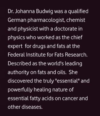
The Interview of Dr Johanna Budwig Omega 3 Lady

Interview by Lothar Hirneise
Lothar Hirneise: What is your fundamental research?
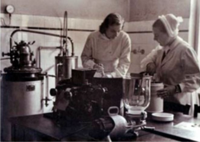
Dr Johanna Budwig: In 1950, I developed Paper Chromatography of fats. With this technique for first time fats, fatty acids and lipoproteins could be detected directly even in 0.1 ml of blood. Using Co 60 I was successful in producing the first differential reaction for fatty acids, and via radioiodine producing the first direct iodine value. I also developed control of atmosphere in closed system by using gas systems which act as antioxidants. Coloring, separating effects of fats and fatty acids were further developed. Behavior was studied in blue light, red light with fluorescent dyes.
I studied the electrical behavior of the unsaturated fatty acids with their “halo” using dyes with rhodamine red. With this technique I proved that electron rich highly unsaturated Linoleic and Linolenic fatty acids (found abundantly in flax oil) are the undiscovered decisive fats in respiratory enzyme function that Otto Warburg could not find. I studied the electromotoric function of pi-electrons of the linolenic acid in the cell membranes, for all nerve function, secretions, mitosis, as well as cell breakdown. I also examined the synergism of the sulfur containing protein with the Pi-electrons of the highly unsaturated fatty acids and their significance for the formation of the hydrogen bridge between fat and protein, which represent “the only path” for fast and focused transport of electrons during respiration.
This immediately caused a furor. Cancer problem was brought in. Hydrogenated fats, which includes all Trans fatty acids proved to be respiratory poisons. This was extensively published in 1950 in many journals including “New Directions in Fat Research.”
Lothar Hirneise: What is the prime cause of Cancer?
Dr Johanna Budwig: In 1928 Dr. Otto Warburg proved that all normal cells have an absolute requirement for oxygen, but cancer cells can live without oxygen – a rule without exception. Deprive a cell 35% of its oxygen for 48 hours and it may become cancerous. Dr. Otto Warburg has made it clear that the root cause of cancer is oxygen deficiency in the cells, which creates an acidic state in the human body.
He also discovered that cancer cells are anaerobic (do not breathe oxygen), get the energy by fermenting glucose producing lactic acid and cannot survive in the presence of high levels of oxygen. Long back in 1911 Swedish scientist Torsten Thunberg postulated that sulfur containing protein (found in cottage cheese) and some unknown fat is required to attract oxygen in the cell. This fat plays a major role in the cellular respiration. For nearly half century scientists were trying to identify this unknown and mysterious fat but nobody succeeded.
Lothar Hirneise: How did you develop cancer therapy which is called Budwig Protocol?
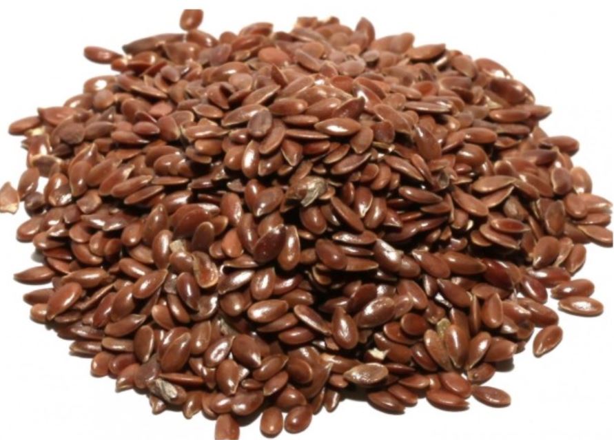
Dr Johanna Budwig: During my research I found that the blood of seriously ill cancer patients was deficient in these unsaturated omega-3 fats (Linoleic and Linolenic fatty acids), lipoproteins, phosphatides, and hemoglobin. In addition, I noticed that cancer patients had a strange greenish-yellow substance in their blood that I could not find in the blood of healthy people. Then I wanted to develop healing program for cancer.
So I decided to straight waygo for human trials and started to give flax oil and cottage cheese to the cancer patients from four big hospitals in Münster. After just three months, patients began to improve in health and strength, the yellow green substance in their blood began to disappear, tumors gradually receded and at the same time as the nutrients began to rise. Thus I had a cure for cancer. It was a great victory and the first milestone in the battle against cancer. My treatment is based on the consumption of flax seed oil with low fat cottage cheese, raw organic diet, detoxification, mild exercise, flax oil massage and the healing powers of the sun. I have treated approx. 2500 cancer patients during last few decades. Prof. Halme of surgery clinic in Helsinki used to keep records of my patients. According to him my success was over 90% and this was achieved in cases where allopathy failed.
Lothar Hirneise: Can you tell us more about the unsaturated fatty acids and their net-like connections?

Dr. Johanna Budwig: Fatty acid is a carboxylic acid having unbranched chain of 4 to 28 carbons. The saturated fatty acids have primarily short carbon chains. In butter, coconut fat, goat fat and sheep fat the fatty acids consists of 4, 6, 8, 10 or 12 carbons, are saturated, however they are also easily co-combustible if the essential fatty acids are present. The unsaturated vital fatty acids really start with the chain with 18 carbon compounds. There are also fatty acids with up to 30 links. Fatty acids with 18 links, like in flax oil with the higher level of unsaturation, are more important for human, particularly for the brain functions of man. Linoleic acid rich in electrons is considered vital. There is a particularly high amount of energy in this double double bond of the linoleic acid.
This energy wanders and is not fixed in place as it is with a chemical compound, such as with table salt. This energy, wandering between electrons and the positively charged protein with sulfur groups is an alternating association process in the electromagnetic field. This is very important. Perhaps you are familiar with the painting of Michelangelo, where God creates Adam (two fingers pointing to each other, however they do not touch). This is quantum physics, the fingers do not touch. The physicists who I know, Max Planck, or Albert Einstein, or Dessauer all represent the view that man is created by God in His image. You see in being together as human beings there is certainly also a connection without directly touching the other person. The dipolarity with a single double bond in olive oil is weaker than it is in sunflower seed oil, which is has two double bonds.
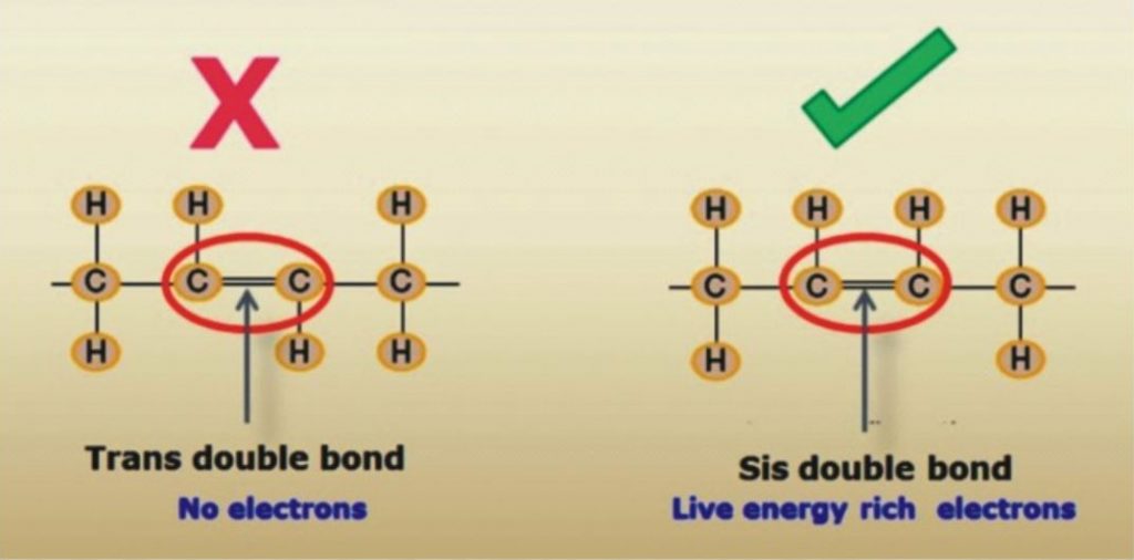
This double double bond is considered to be vital for man. However if the same chain length of 18 carbons has three unsaturated fatty acid compounds, then the electrical energy is as strong as a magnet. This electronic energy is negatively charged. The positively charged sulfur groups of the protein adhere in the unsaturated bonds where the electrons are and that is where they insert their sulfur-containing compounds.
This produces the lipoproteins. The life process is sustained in the interplay between the positively-charged particles and negatively-charged particles. In this process there is no connection, and this is our life element. If radical damage occurs at this point through fatty acids that has lost electron energy, but rather are cross-linked like a net, then the dipolarity can no longer work actively in this net. This is the deadly effect of radicals, because instead of the chains with the electron clouds they interlace a net without electron clouds, indeed with unsaturated bonds, but without dipolarity. I quickly knew that the triple unsaturated fatty acids, which were called linolenic acid, and which no one had isolated before me, had 18 links and that they did not always carry their double bonds at the same point. They have such a strong electronic energy compared to the heavier matter in the 18-link fatty acid chains, that biologically this energy is far greater than it is with the next arachidonic acid with 20 links. The highest electron collection is with the combination of linoleic-linolenic fatty acids in flax oil. The linolenic acid as conjugated (interaction of neighboring double bonds in the molecule that are separated by a single bond) fatty acid is even more effective and is even more strongly interplay with linoleic acid as it is present in the flax oil for oxygen absorption. This was relatively easy for me to verify in my experiments. I would like to emphasize this. The combination of double unsaturated linoleic acid with triple unsaturated linolenic acid is particularly well-combined in flaxseed.
Lothar Hirneise: Is it this energy that heals cancer?
Dr. Johanna Budwig: Yes, this energy is now movable and it is easily released. It is precisely this energy that heals cancer, or does not even allow it to occur. If this vital element is present then no tumor can exist. This vital element is a deciding factor in the immune system. There is no effective factor in the immune system other than the essential fatty acids.

Lothar Hirneise: What is an electron cloud?
Dr Johanna Budwig: If the enhancement of electronic energy is always higher through absorption of sun photons in the unsaturated fatty acids e.g. in linolenic fatty acids, then the power of the electrons is so high in the dipolarity in between gravity and electrons, that they lifts off of the heavy mass and floats like a cloud hence I called them electron cloud.
Lothar Hirneise: What is the significance of the cloud?
Dr. Johanna Budwig: No life form has as much energy to store the electrons and photons as does man. The electronic energy stored particularly in the vital, highly unsaturated fatty acids, is very strong life element for man. Man cannot live without them. If oils are treated with heat and harsh chemicals (during refining and hydrogenation process to increase their shelf life) then the wealth of vital electronic energy is destroyed and Trans fats are formed with net like connections. They are no longer vital fats with 18 links, but rather they form cross-links between the fatty acids like a large net, and are highly damaging to our body, do not adhare with proteins, do not attract oxygen and act like a radicals. I repeat because it is so important: I have detected particles in oils treated with steam, which indeed have a positive iodine value, but which are highly toxic for man.
Lothar Hirneise: So you preach against these toxic hydrogenated and refined oils?
Dr. Johanna Budwig: I am completely against using these “pseudo” fats – “hydrogenated”, “partially hydrogenated”. These are the biggest enemy of mankind. I had scientific proofs. The heart rejects these fats and they are deposited as inorganic fat on the heart muscle itself. They end up blocking circulation, damage heart action, inhibit cell renewal and impede the free flow of blood and lymph fluids.
But it was highly profitable business for multinationals. When I preached against these fats, they stood against me, first they tried to give me hush money and when I refused they filed many fake court cases against me. I was working for humanity and had scientific proof.I was like rock of Gibraltar in my decision; I fought and won all the cases ultimately.
Lothar Hirneise: What is mysterious marriage of sun and moon in terms of quantum physics?
Dr. Johanna Budwig: The photon, the quantum, is the smallest component of the sunlight. It is the fastest moving traveler, it speeds along with time. The photon is always in motion. Nothing can ever halt its motion. The photon is full of all colors, and can change its color and its frequency. The photon is the purest form of energy, the purest wave, can unite with a second photon when it is in resonance with the other, to form a “short-lived particle.” This particle, known as a p0 particle, can break up into two photons again, without mass, as a pure wave in motion. This is the basis for the wonderful back and forth movement between light and matter. The photon can never be pinned down to one location. This is the foundation for the Theory of Relativity.
This very active, dynamic and energetic photon can be captured by electron that are in resonance with the photon. What does that mean? Electrons are already a component of matter. They are also continuously in motion. They constantly oscillate on their own wavelength. They have their own frequency, just like a radio receiver that is set to a specific wavelength.
The electron loves photons. It attracts photons by its magnetic field. When an electrical charge moves, it always produces a magnetic field. The moving photon also has a magnetic field. Both fields attract each other when the wavelengths are in tune. The wave length of the photon – which the photon can change – must fit into the wavelength of the orbiting electron so that the orbit maintains a complete wavelength. This is eternal love between sun-god, photons and moon-goddess, electrons, this is the mysterious marriage of the sun and moon in terms of modern science.This feature is extremely interesting in terms of its physical manifestation, its biological and even its philosophical consequences. All matter has its own inherent vibration. Of course, this also applies to living substances. The energy which is being absorbed must correspond to the inherent wavelength.
The sun rays are very much in harmony with humans. It is no coincidence that we love the sun. The resonance in our biological tissue is so strongly tuned to the absorption of solar energy that there is nothing on earth that has a higher concentration of solar energy photons than humans. This enrichment with solar energy depends strongly on the “like energy” aspects, a wavelength that is compatible with humans, and this is supported when we eat foods that have electrons with electromagnetic fields that attract the photons of the sun. An abundance of these electrons, which are tuned to the solar energy frequency, exist, in many seed-oils. Scientifically these oils have even been designated as electron-rich, “essential,” highly unsaturated fats. But when we began processing fats to increase the shelf life, nobody thought about the significance that this would have for the survival and the further development of the human species. We destroyed their extremely important wealth of electrons, which are very mobile and react so wonderfully to sunlight.
Lothar Hirneise: What is the concept of “human” and “anti-human” in terms of quantum physics?
Dr. Johanna Budwig: It is interesting that in the science of quantum physics the concept of “human” has already been coined. It is “human”, with the highest accumulation of photons, always striving toward the future, who possesses within himself the highest potency of solar energy on the earth. The physicists project from mathematical equations that man, with his wealth of electrons, is directed forwards future in time.
Using mathematical equations that are valid in physics, reversing the time quotient represents the mirror image of man — the “anti-human,” whereas “man” represents the picture of highest rank in terms of physics, i.e. directed against entropy. The “anti-human” is directed back in time. The “anti-human” possesses few solar energy photons, in physical terms electron-poor, directed into the past — also in his thinking — is paralyzed in his life functions, lacks energy and strength because he is missing the electrons that are in harmony with the sun as “life-element.”
The physical processes which are generated through the use of X-rays, gamma rays, atom bombs, or cobalt radiation, are pointed in the same direction as the development toward the “anti-human,” from the perspective of physics and mathematics. The electron structure of the life functions is destroyed by these rays. According to the so-called “World Line” and the Theory of Relativity of modern physics, time and space are connected together in one equation.
It is very interesting to investigate our food from this perspective. Fats that have had their electron structure destroyed to make them keep longerpromote the development of the “anti-human”and have a very detrimental effect on the future-directed, electron-rich human being, according to the “World Line diagram.” They disturb the electron exchange within living tissue because they, like tar, act as insulators against electrical conductivity, plainly deaden the life functions at the respective operative locations, e.g. in organs, and in growth centers of the body, as well as throughout the whole body. They also promotes cancer.
On the other hand, electron-rich nutrition, electron-rich highly unsaturated oils,herbs and fruits which are rich in aromatics and natural color components that correspond to the colors of the photon s of sunlight — all these increase the absorption, storage and utilization of the sun’s energy.
Lothar Hirneise: What is your view point about surgery for tumors?
Dr. Johanna Budwig: I am totally against radiation and chemo; I also reject hormonal treatment. Surgery must be considered individually. I am not a proponent of quickly making artificial anus. Conventional oncology no longer does justice to the cancer patients.
Lothar Hirneise: You also studied medicine at the age of 47 years.
Dr. Johanna Budwig: (smiling) Yes handsome! That’s right, my opponents were accusing me that how can I treat cancer patient without doctors degree. This thing pinched me, so in 1955,I joined medical school in Göttingen. There I was using my therapy very successfully in various clinics. I still remember the time I was working late one night in Göttingen, a woman came to me, with her small child whose arm was supposed to be amputated due to a tumor. I treated her and soon the subject of amputation was dismissed and the child quickly did very well.
Because I was still a medical student at this time, I was summoned to appear before the Municipal Court due to a petition that I should be prohibited from studying medicine. I explained the truth in the court. The judge rejected the case and said, “You have done a good job. In my area of jurisdiction nothing will happen to you. If it does there will be a scandal in the scientificcommunity.”
Lothar Hirneise: What do you recommend for prevention of cancer?
Dr. Johanna Budwig: Only flax oil as oil. I reject frozen and preserved meat. Fresh meat is OK. No frozen food and no bakery products. Oleolux should be used as butter. Prepare fruit juices yourself. Cheese and potatoes are OK. Also the electromagnetic environment (e.g. microwave and mobile phones etc.) in which we live is very important. I reject synthetic textiles and foam mattresses because they steal lot of forces from you. A lot of wood in home construction and woolen or silk carpets are also important. Wear gemstones, they also have good biological radiation. The environment and living conditions must be as biological (organic & natural) as possible. Regular sleep is very important.

pH Vs Coronavirus
Here are some excerpts of a paper on the functioning of Hydroxychloroquine, the drug being touted as a medicine to combat CoronaVirus. It can be seen that it’s major action, supportive of the previous data I’ve presented, is raising pH…
Hydroxychloroquine (HCQ) enters and accumulates in lysosomes along a pH gradient. In lysosomes, hydroxychloroquine inhibits the degradation of cargo derived externally (via endocytosis or phagocytosis) or internally (via the autophagy pathway) in autolysosomes by increasing the pH to prevent the activity of lysosomal enzymes. Inhibition of lysosomal activity can prevent MHC class II-mediated autoantigen presentation.
Hydroxychloroquine can also accumulate in endosomes and bind to the minor groove of double-stranded DNA. This drug can inhibit Toll-like receptor (TLR) signalling by altering the pH of endosomes (involved in TLR processing) and/or preventing TLR7 and TLR9 from binding their ligands (RNA and DNA, respectively). Hydroxychloroquine can also inhibit the activity of the nucleic acid sensor cyclic GMP-AMP (cGAMP) synthase (cGAS) by interfering with its binding to cytosolic DNA. By preventing TLR signalling and cGAS–stimulator of interferon genes (STING) signalling, hydroxychloroquine can reduce the production of pro-inflammatory cytokines, including type I interferons.
….
As the pH in lysosomes is optimal for lysosomal enzymes involved in hydrolysis, by increasing the pH of endosomal compartments85, chloroquine and hydroxychloroquine might impair the maturation of lysosomes and autophagosomes and inhibit antigen presentation along the lysosomal pathway (Fig. 3).
…
These processes possibly occur, at least in part, through drug-mediated changes in the pH of autophagosomes and/or lysosomes, which indirectly influence immune activation; however, such a mode of action requires additional validation to aid with future drug development.
https://www.nature.com/articles/s41584-020-0372-x
This is the drug they are saying shows great promise for Coronavirus treatment.
Here is excerpts from a paper just being released, where studies have shown 100% cure rate with CoronaVirus:
He talks about lowering the acidity in the cells…
https://www.covidtrial.io/
From their prelim paper:
Specifically, the CDC research was completed in primate cells using chloroquine’s well known function of elevating endosomal pH. The results show that “We have identified chloroquine as an effective antiviral agent for SARS-CoV in cell culture conditions, as evidenced by its inhibitory effect when the drug was added prior to infection or after the initiation and establishment of infection. The fact that chloroquine exerts an antiviral effect during pre- and postinfection conditions suggest that it is likely to have both prophylactic and therapeutic advantages.
…
When chloroquine is added after infection, it can rapidly raise the pH and subvert on-going fusion events between virus and endosomes, thus inhibiting the infection. When added after the initiation of infection, it likely affects the endosome-mediated fusion, subsequent virus replication, or assembly and release. Specifically, rapid elevation of endosomal pH and abrogation of virus-endosome fusion may be the primary mechanism by which virus infection is prevented under post-treatment conditions.
…
Specifically, the virus depends on turning over the host proteins to trigger response for available building blocks to make their own proteins or nucleic acids. They break down due to low PH catalyzed by hydrolysis.
…
It is this part of the coronavirus’ replicative path that chloroquine inhibits. Notably, because of its nitrogen structure, chloroquine has the unique ability to get into cells and cross endosomal membranes. Once inside, nitrogens in chloroquine (and quinines in general) prevent acidification by absorbing a high amount of hydrogens that simply then interact with nitrogen and then chloroquine becomes positively charged – an ionic interaction which makes it harder for the endosome to become acidified. The result is a buffer that holds it at the higher pH and prevents it from becoming acidic enough to be functional. To summarize, because chloroquine has a multitude of extra nitrogens, once it crosses the membrane and enters an organelle, the organelle is prevented from reaching a lower pH. The organelle’s enzymes cannot work because the donor group will be a hydrogen ion, disabling the hydrolysis required for coronavirus replication. This means that all kinds of events in the cell are incapable of performing optimally, including viral replication.
https://github.com/covidtrial/info/raw/master/An%20Effective%20Treatment%20for%20Coronavirus%20(COVID-19).pdf
Biological Ionisation…
Wonder why I go on and on about pH…
Stunning nobody is talking about Milk Of Magnesia, or other inexpensive pH raising substances…
Related:
Bill Munro Vs The Coronavirus
Bill brought inhalation of H2O2 to the publics attention, he used it 5-6 pumps, 6 or 7x a day. To keep his Oxygen levels high, to keep the Viruses out, which come when Oxygen is down.
This was something I tried many years ago, but didn’t feel a direct or urgent need to do, so put it into my memory locker.
However, after studying this new Viral Epidemic which increasingly surrounds us, I pulled the information back to my short term memory to mix with the pH principles I had been reflecting on.
My daughter has suffered from Chronic Lung Disease as a result of being born at 24weeks gestation, so Oxygen and Viral susceptibility has been something I am used to reflecting deeply upon.
Anyway, the data shows that if you are of a lower pH, this virus group (Corona) is 10x more forceful and you are 10x more susceptible (at a pH of 6 Vs 7), so after ensuring I got my diet plans and supplements into order, I began to reflect over and over how else I can best prepare.
This is when Bill Munro came back to my mind, so I ordered 2x Nasal Vaporiser’s for about £3, and I decided that I would start taking a similar number of rounds of H2O2 as Bill did, but diluted to 1.5% with 50% Distilled water and 50% Food Grade H2O3 at 3%.
The literature for killing the Virus shows that 0.5% H2O2 kills the virus in 1 Minute, so 1.5% should be effective in 20 Seconds or thereabouts. So, to my unqualified mind, this leads me to presume that several rounds of this daily may safeguard me against this threat, if not preventing it from entering my airways entirely, then at least killing it off 5-6 times a day, minimising the intensity of the infection as best I may.
When I have talked of this method to others, peoples minds immediately seem to jump to “you are swallowing bleach”, with a shocked reaction, yet I explain that this is simply water with an extra Oxygen molecule attached, which people regularly treat their teeth with at 7% some 450% stronger than I am using. People seem to not bat an eyelid at using 4.5x stronger bleach for their teeth and vanity, yet to stave of seemingly very harsh virus people react like I am the crazy one!
This method along with Alkalising my terrain via diet and supplements are the best methods I have encountered to protect myself against this horrid viral outbreak.
If the outbreak worsens in the UK, I am intending to upgrade to the full 3%
Thank you Bill.